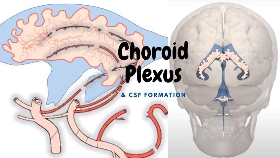
What is choroid plexus ?
As we know, the blood vessels supplying the brain are present within subarachnoid space (between arachnoid mater & pia mater).
During the developmental process, in the ventricular system of the brain, the ventricular wall is so thin that artery protrude out into the ventricular system along with piamater
Thus the double folding of piamater enclosing tufts of capillaries is called tela-choroidea.
Such fold of piamater is lined by secretory ependyma cell which is collectively called as choroid plexus.
CSF from the choroid plexus of the lateral ventricles is derived from the plasma filtration of blood capillaries which are from the anterior choroidal artery.
Similarly, the choroid plexus of the fourth ventricle is traversed by anterior inferior cerebellar artery & superior cerebellar arteries, branch of basilar artery & posterior inferior cerebellar artery, branch of the vertebral artery.
Layers of choroid plexus from inner to outward
1. Ependymal cells
2. Fold of Piamater
3. Endothelial cells of blood capillaries
You can learn more:
How CSF is formed from the choroid plexus?
(In a simple description)
CSF is formed by an active transport mechanism followed by a passive diffusion process of blood plasma present in the blood capillaries of choroid plexus.
The ependymal cell has Active Na+ transporter. These transporters with the active utilization of ATP, actively transport Na+ into the ventricular system. Similarly, it is followed by Chloride (Cl-). Thus this process increased the osmolarity pressure within the ventricular system. Thus there is possible of passive diffusion of water from the blood capillaries of the choroid plexus into the ventricle.
Similarly, there is the presence of glucose transporter called GULT-1.
As we know, GLUT-1 is glucose-dependent, not the insulin-dependent transporter.
GLUT-4 which is found in muscle & adipose tissue is only insulin-dependent as without insulin glucose cannot enter into the cell.
Anyway, the level of insulin doesn’t affect the transport of glucose in the brain.
With the help of GLUT-1 transporter, glucose is also transported into the ventricle forming one of the compositions of CSF.
Thus, CSF also helps to nourishes the nervous tissue. Only CSF comes directly in contact with neurons. Even blood directly can’t come in contact with neurons of the brain. It provides nutrition to neurons & returns waste metabolic products to venous sinuses.
Thus, to sum up, as a whole, CSF is formed by selective transport mechanism of blood plasma whose composition is quite similar to that of blood plasma except CSF is somehow protein less or very low protein content.
Tentative Comparison between CSF & Blood plasma
| Components |
CSF |
Blood Plasma |
| Water (%) |
99 |
92 |
| Sodium (meq/L) |
138 |
138 |
| Potassium (meq/L) |
2.9 |
4.8 |
| Chloride (meq/L) |
119 |
102 |
| Protein (mg/dl) |
20 |
7000 |
| Glucose (mg/dl) |
60 |
95 |
| Osmolarity (mOsm/L) |
295 |
295 |
| pH |
7.33 |
7.4 |
| PCo2 |
50 |
39.5 |
Circulation of CSF
CSF formed by Choroid Plexus of Both left & right lateral Ventricles
↓
Two respective intraventricular foramen
↓
Third Ventricles
(Hypothalamus forms the floor & lateral wall below the hypothalamic sulcus of Third Ventricle)
(Remaining lateral wall of the upper part of the Third Ventricle is formed by Thalamus)
↓
Aqueduct of midbrain
↓
Fourth Ventricle
↓
From 4th Ventricle below, there are four major openings for the circulation of CSF.
1. Straight down continuation of 4th ventricle - as Central canal of Spinal Cord
2. One median opening on the back.
3. Two lateral openings on either side.
↓
From 1 median & 2 lateral openings
↓
Opens in
1. Subarachnoid space around spinal cord & cauda equina
↓
Absorbed by veins of spinal cord
2. Cerebellomedullary cistern & pontine cistern, (i.e. they are subarachnoid spaces in cranial cavity)
↓
Tentorial notch
↓
↓
↓
Arachnoid granulations
(Projection of arachnoid member into dural sinuses for passage of CSF from subarachnoid space into venous system)
↓
Absorption
↓
Superior sagittal sinus
Comments (0)