Cerebrum:
Location:
- Anterior Cranial Fossa
- Middle Cranial Fossa &
- Supratentorial part of Posterior cranial fossa
Note: Below the tentorium cerebelli lie cerebellum in posterior cranial fossa.
Features
* Cerebrum has two cerebral hemispheres.
* Both hemispheres are incompletely separated by median longitudinal fissures.
* Because below, both hemispheres are connected to each other by white matter called corpus callosum (a type of commissural fibers).
* Each hemisphere contains a cavity called the lateral ventricle.
Note:
| Contents of median longitudinal fissure is falx cerebri with superior sagittal sinus and inferior sagittal sinus. |
| The main composition of white matter is nerve fibers/ tract. |
| Corpus callosum (white matter) is the compact bundle of nerve fibers / consisting of millions of axons connecting two hemispheres. |
Tips:
| Nerve fibers are axons or dendrites of the nerve cell body. |
|
The tract is the bundle of nerve fibers.
Thus, tract and nerve fibers are called white matter.
|
External Features of the cerebral hemisphere:
Three surfaces:
1) Superolateral surface
* Related to the cranial vault
2) Medial surface
* Present on either side of the median longitudinal fissure
3) Inferior surface - divided into two parts by lateral sulcus
* Orbital part and Tentorial part of the inferior surface
Note: Tentorial part of the inferior surface rest on the Tentorial cerebelli.
Four Borders
1) Superomedial border
Separate the superolateral surface from the medial surface
2) Inferolateral border
Separate the superolateral surface from the inferior surface
3) Medial orbital border
Separate the medial surface from the orbital part of the inferior surface
4) Medial occipital border
Separate the medial surface from the tentorial part of the inferior surface.
Three poles (Pointed end):
- Frontal pole
- Temporal pole
- Occipital pole
Fours lobes of the Cerebral Hemisphere:
Frontal lobe:
Important for voluntary motor functions, motivation, emotions, affect, and awareness of self. |
Parietal lobe:
A major center for interpretation of all sensory information except smell, hearing, and vision. |
Occipital lobe:
Center for the integration of visual input. |
|
Temporal lobe:
The center for smell and hearing plays an important role in memory.
|
How lobes are divided?
- Central sulcus
- Lateral sulcus
- 1st & 2nd imaginary lines
* Central sulcus divides frontal and parietal lobes.
* Lateral sulcus divides the frontal & temporal lobes.
* 2nd imaginary line which is drawn as the continuation of the posterior ramus of the lateral sulcus divides the parietal lobe & temporal lobe.
* 1st imaginary line drawn between parieto-occipital sulcus and pre-occipital notch separate occipital lobe from parietal & temporal lobe.
Note:
The elevated part is known as the gyrus.
The depressed part is known as the sulcus.
The cerebral cortex is folded into gyrus which are separated from each other by sulci.
Central Sulcus:
The central sulcus begins at the superomedial border, 1.25cm behind the midpoint of the frontal pole and occipital pole.
* Then it course obliquely downwards & forward.
* And end a little above the posterior ramus of the lateral sulcus.
Note:
| The central sulcus extends for a short distance onto the medial surface. |
|
And the U-shaped gyrus around the central sulcus is called the paracentral lobule [on the medial surface].
|
Lateral Sulcus:
The lateral sulcus begins on the inferior surface.
* It separates the inferior surface of the cerebrum into the orbital part and the Tentorial part.
Laterally, this sulcus reaches the superolateral surface, it is divided into three ramus.
- Anterior horizontal ramus
- Anterior ascending ramus
- Posterior ramus
The posterior ramus extends backward and slightly upward over the superolateral surface.
Parieto-occipital sulcus:
Parieto-occipital sulcus is the sulcus of the medial surface.
Its upper end shortly cut the superomedial border about 5cm in front of the occipital pole.
Preoccipital notch:
This notch is present on the inferolateral border about 5cm in front of the occipital pole.
- 1st imaginary line [on the superolateral surface] is drawn between the parieto-occipital sulcus & pre-occipital notch.
- 2nd imaginary line is drawn as the continuation of the posterior ramus of the lateral sulcus up to 1st imaginary line.
Note:
| Central sulcus and lateral sulcus are the main sulcus of the superolateral surface. |
| But parieto-occipital sulcus is the sulcus of medial surface. |
|
The content of the lateral sulcus is the middle cerebral artery which is the continuation/ branch of the internal carotid artery.
|
Functional classification of Sulcus:
- Limiting sulcus (Eg. Central sulcus)
- Axial sulcus (e.g Postcalarine sulcus)
- Operculated sulcus (Eg. Lunate sulcus)
Limiting sulcus
It separates two areas that are functionally and structurally different.
Example: Central sulcus
Limiting the sulcus, as it limits the motor area from the sensory area.
* In front of the central sulcus, is the motor area.
* Behind the area of the central sulcus is the sensory area.
| Anterior to it lies motor cells that initiate the movements of the opposite side of the body. |
|
Posterior to lies the general sensory cortex receives sensory information from the opposite side of the body.
|
Simply,
1) Frontal lobe - motor in function
2) Parietal lobe - mainly area of sensory impulse integration
3) Temporal lobe - for the sense of hearing
4) Occipital lobe - the sense of vision
Sulci and gyrus of the cerebrum:
Note:
The central sulcus and lateral sulcus are not the sulci of the particular lobe.
They act as a partition between the lobes.
Note:
Anterior horizontal and anterior ascending rami of the lateral sulcus subdivide the inferior frontal gyrus into three parts:
Pars orbitalis
Pars triangular
Pars opercular's
The area behind the postcentral gyrus is divided into the superior and inferior parietal lobules by the intraparietal sulcus.
The calcarine sulcus starts below the splenium and courses forward toward the occipital pole.
It gives off the parieto-occipital sulcus which runs toward the superolateral surface.
The visual cortex lies above & below this sulcus.
The cingulate gyrus lies between the corpus callosum and the cingulate sulcus.
Frontal lobe:
The boundary of the Frontal Lobe:
- Superiorly - Superomedial border
- Inferiorly - lateral sulcus
- Posteriorly - central sulcus
Functional areas of the Frontal Lobe:
|
Primary motor area (area no.4)
|
|
Located in the precentral gyrus & extends up to the anterior part of the paracentral lobule.
* About 30% of the corticospinal tract & corticonuclear tract (Pyramidal Tract) arise from here.
Control the voluntary activities of the opposite half of the body
|
The lesion in the motor area:
Contralateral paralysis (Hemiplegia of extremities of the opposite side)
|
|
Premotor area (area no.6)
Control extrapyramidal system
* Needed for perfect voluntary motor movements initiated by the Primary motor area
|
The lesion in the premotor area
Contralateral paralysis
* Difficulty in the performance of skilled movements
|
|
Frontal eye field (area no.6,8)
Controls horizontal conjugate movements of the eyes
|
The lesion in frontal eye field
Horizontal conjugate movements of the eyes are lost
|
|
Motor speech area/ Broca’s area (area no.44,45)
Control spoken speech
|
|
Lesion of Broca’s motor area
Aphasia (motor)/ inability to produce speech
|
Pre-frontal area (area no.9,10,11,12)
Controls emotion, concentration, attention, & judgment.
|
|
The lesion in the pre-frontal area - Loss of orientation
Abnormality in ideas, judgment, emotional feeling & personality.
|
Parietal lobe:
The boundary of the Parietal Lobe:
- Anteriorly - Central sulcus
- Posteriorly - 1st imaginary line from preoccipital notch to parieto-occipital sulcus
- Superiorly - Superomedial border
- Inferiorly - 2nd imaginary line drawn as the continuation of the posterior ramus of the lateral sulcus
Functional areas of the Parietal Lobe:
Primary somatosensory area (area no.3,1,2)
Situated in the postcentral gyrus & extends up to the posterior part of the paracentral lobule
Primary Center for Exteroceptive Sense (Pain, Temperature, Pressure, Touch)
* and Proprioceptive Sense (Movements of Joints, muscles, Vibration) from the opposite side |
Lesion:
Loss of Proprioceptive & Exteroceptive sense from the opposite side. |
Sensory association area (area no.5,7)
Responsible for the Prediction of the shape, size, and structures of objects.
One can recognize what has been placed in their hands without seeing.
This capability of a person is called stereognosis.
|
Lesion:
Loss of stereognosis |
|
Wernicke’s area/ Sensory speech area (area no.22)
Responsible for understanding the spoken speech
|
Lesion:
Sensory Aphasia |
Note:
Aphasia means impairment of language, affecting the production or understanding(sense) of speech and the ability to read & write.
It could occur due to stroke, or brain tumor. |
|
Broca’s motor speech area lesion:
One loses the ability to produce proper speech. A person finds difficulty in expressing words he/she wants, but could understand the speech.
|
|
Wernicke’s sensory speech area:
Loss of ability to understand the speech & written word, but can speak fluently.
|
Temporal lobe:
The boundary of the Temporal Lobe:
- Superiorly- Posterior ramus of the lateral sulcus
- Inferiorly - Inferolateral border
- Anteriorly - The temporal pole
- Posteriorly - 1st imaginary line from preoccipital notch to parieto-occipital sulcus
Functional Areas of Temporal Lobe
Primary Auditory Area (area no. 41, 42)
Reception & Perception of Auditory Impulses |
Lesion:
Impaired Hearing |
Secondary Auditory Area (area no. 22)
Correlate the auditory impulses to the past memory |
Lesion;
Auditory Agnosia
(Lost of past auditory memory)
One cannot recognize or identify the sound. |
Occipital lobe:
Boundary of occipital lobe
- Superiorly - superomedial border
- Inferiorly - Inferolateral border
- Anteriorly - 1st imaginary line from preoccipital notch to parieto-occipital sulcus
- Posteriorly - Occipital lobe
Functional areas of the Occipital Lobe
Primary visual area (visuosensory area) - (area no.17)
Receives and integrates the visual impulses |
Lesion:
Homonymous Hemianopia
(Defect in nasal field of vision of one eye & Temporal field of vision of another eye) |
Secondary visual area (area no.18,19)
Correlate the recent visual impulses with the past memory.
|
Lesion:
Visual Agnosia
(Lost of past Visual memory)
One cannot correlate the visual impulses with a past memory, so fail to recognize the seen objects. |
Paracentral lobule:
It is the U-shaped gyrus on the medial surface around the extended end of the central sulcus.
It is the lobe on the medial surface of the cerebral hemisphere.
Subdivision:
The central sulcus extends shortly on the medial surface and divides the paracentral lobule into:
- Anterior part - continuous with precentral gyrus of the frontal lobe
- Posterior part - continuous with postcentral gyrus of the parietal lobe
Function of paracentral lobule:
- Control the motor and sensory innervation of the opposite side of the lower extremities.
- It is the chief center for micturition & defecation.
Anterior part -
* Regulates movement of the contralateral lower extremity
* Voluntary control of micturition & defecation
Posterior part -
* Receives sensation from the contralateral lower extremity.
* Sense of distention from bladder & rectum.
Note
| Micturition is the parasympathetic function. |
|
The lower spinal level Center of micturition is located on the S2-S4 segment of the spinal cord and parasympathetic fibers are carried by pelvic splanchnic nerves.
|
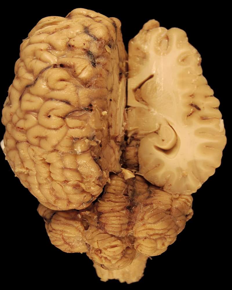
The white matter of cerebrum/ Fibers of Cerebrum:
The main component of the white matter of the cerebrum is myelinated nerve fibers
Subdivision of white matter
- Association (Arcuate Fibers)
- Commissural Fibers
- Projection Fibers
Association (Arcuate Fibers):
Fibers that connect different areas of the same hemisphere
Two types:
1) Short association fibers
* These fibers connect the adjacent area of the same lobe
2) Long-association fibers
* Connect distant areas of different lobes of the same hemisphere.
Examples
- Uncinate Fasciculus
- Cingulum
- Inferior longitudinal Fasciculus
- Superior longitudinal Fasciculus
Note
|
Superior longitudinal Fasciculus:
Connect occipital lobe with frontal lobe; form pathway of accommodation reflex; occipital cortex to frontal eye field
|
Commissural fibers:
Fibers that connect corresponding parts of two hemispheres.
Example
- Corpus callosum
- Anterior commissure
- Posterior commissure
- The commissure of the fornix (hippocampal commissure)
- Habenular commissure
Corpus Callosum:
The Corpus callosum is the largest commissural fiber of the brain.
* The central part of the medial aspect of the hemisphere is occupied by the corpus callosum.
* It is made up of nerve fibers connecting the two cerebral hemispheres.
Parts of the corpus callosum:
- Rostrum
- Genu
- Trunk/Body
- Splenium (Posterior end)
The function of the corpus callosum:
Responsible for the transfer of information that is essential for the bilateral response.
Tips
Below the corpus callosum, there are the septum pellucidum, the fornix, and the thalamus.
* Lateral to septum pellucidum, lies the central portion of the lateral ventricle.
Projection Fibers:
Connect the cerebral cortex to other parts of CNS like the brainstem, spinal cord
Example
- Internal capsule
- Corona Radiata
- Optic radiation
- Fornix
Internal capsule:
The internal capsule is the compact bundle of nerve fibers (a part of white matter) present between the thalamus & lentiform nucleus.
* It consists of ascending and descending nerve fibers that connect the cerebral cortex to the spinal cord & brain stem.
* Almost all fibers going to the cerebral cortex or fibers coming from the cerebral cortex, all pass through internal capsules.
Continuation:
* Upward with corona radiata
* Downward with crus cerebri of midbrain
Division of internal capsule:
- Anterior limb
- Genu
- Posterior limb
- Retrolentiform part- lies behind the lentiform nucleus
- Sublentiform part - lies below the lentiform nucleus
The boundary of internal capsule:
* Medially- Head of caudate nucleus & thalamus
** Laterally- Lentiform nucleus
Artery supply of internal capsule:
Anterior limb by:
* Recurrent branch of anterior cerebral artery
* Direct brach from anterior cerebral artery |
Genu by
* Direct branch from the internal carotid artery
* Branches from the posterior communicating artery |
|
Posterior limb by:
* Medial & lateral striate arteries of the middle cerebral artery
|
Sublentiform part & retrolentiform part by:
* Branches from the posterior cerebral artery |
Fibers of Internal capsule
| Parts |
Motor fibers |
Sensory fibers |
| Anterior limb |
Corticopontine fibers |
Anterior thalamic radiation |
| Genu |
Corticopontine fibers
Corticospinal fibers
|
Superior thalamic radiation |
| Posterior limb |
Corticopontine fibers
Corticospinal fibers |
Superior thalamic radiation |
| Retrolentiform part |
Corticopontine fibers |
Optic radiation |
| Sublentiform part |
Corticopontine fibers |
Auditory radiation |
Note:
All parts of the internal capsule contain corticopontine fibers.
** Because of widely pass of motor and sensory nerve fibers within the internal capsule, even a small lesion in the internal capsule could cause paralysis and sensory loss in the opposite half of the body.
Clinical importance of internal capsule:
The lesion of the internal capsule is usually vascular.
* Posterior limb of the internal capsule is supplied by lateral & medial striate branches of the middle cerebral arteries. These are end arteries.
* These arteries are vulnerable to rupture leading to loss of sensation & spastic paralysis of the opposite half of the body (contralateral hemiplegia).
* It comes under the category of upper motor neuron type of paralysis.
Artery supply of Cerebrum
According to surfaces :
On the superolateral surface:
The main artery is the middle cerebral artery, (continuation or terminal branch of the internal carotid artery)
|
The superior part of the superolateral surface extending 2.5cm width from the superomedial border up to the parietals-occipital sulcus
* Supplied by anterior cerebral artery
|
|
The lower part of the temporal lobe (excluding the temporal pole) and occipital lobe
* Supplied by posterior cerebral artery
|
|
The rest of all (maximum) areas
* Supplied by the middle cerebral artery.
[Including Broca’s area, Wernicke’s area, and temporal pole.]
|
On the medial surface:
The main artery is the anterior cerebral artery, a terminal small branch of the internal carotid artery
Temporal pole of the temporal lobe
* Supplied by middle cerebral artery |
Rest of the temporal (excluding the temporal pole) & occipital lobe
* Supplied by posterior cerebral artery |
|
Rest of the upper maximum part area including the paracentral lobule
* Supplied by anterior cerebral artery
|
On the inferior surface:
The main artery is the posterior cerebral artery, a branch of the basilar artery.
Most parts of the temporal lobe (except the temporal pole) & occipital lobe
* Supplied by posterior cerebral artery |
Temporal pole & lateral part of the orbital surface of the frontal lobe
* Supplied by middle cerebral artery |
|
A medial small part of the orbital surface of the frontal pole
* Supplied by the anterior cerebral artery.
|
Tips:
Always remember
Temporal pole - by middle cerebral artery
* Occipital lobe - by posterior cerebral artery
Note:
The course of the anterior cerebral artery
Anterior cerebral artery runs along above the course of the corpus callosum on either side of the median longitudinal fissure - middle surface. |
The course of the middle cerebral artery
Middle cerebral artery course along the lateral surface which begins from the inferior surface and runs laterally in the superomedial surface and runs along the posterior ramus of the lateral sulcus. |
| Content of lateral sulcus: Middle cerebral artery |
Lateral sulcus - starts from the inferior surface
* Divide the inferior surface into the orbital part & Tentorial part
* On coming on superolateral surface
The lateral sulcus give 3 rami:
1) Anterior horizontal ramus
2) Anterior ascending ramus
3) Posterior ramus |
Anterior horizontal & ascending rami divide the inferior frontal gyrus into:
a) Pars triangularis
b) Pars opercularis
c) Pars orbitalis |
|
Location of Broca’s motor speech area:
I) Pars triangularis (area no .45)
II) Pars opercularis (area no. 44)
|
Stroke is related to the brain, not the heart.
Histology of Cerebrum:
The cerebrum consists of outer grey matter knowns as the cerebral cortex & inner white matter made of bundles of nerve fibers.
Within the white matter of its basal parts, the embedded mass of grey matter is found called Basal Nuclei.
The difference in the arrangement of White matter & Grey matter In the Brain & Spinal Cord
Both the spinal cord & brain contains grey and white matter.
In Brain
Cerebrum:
Outer cortex of Grey matterInner core of White matter |
Brainstem(Midbrain, Pons, Medulla)
Outer White matter
Within the white matter, embedded masses of Grey matter are called nuclei.
Numerous nuclei are present in the brainstem within the white matter. |
In Spinal Cord
Outer white matter
Inner H-shaped Grey matter
Note:
The grey matter consists of nerve cell bodies & dendrites.
White matter consists predominantly of myelinated nerve fibers.
Development of cerebrum:
Simply, the Brain & Spinal cord develops from Neural Tube.
The cranial part of the neural tube differentiates into 3 main enlarge vesicles.
| Parts |
Subdivision |
|
Forebrain
(Prosencephalon)
|
Telencephalon(Cerebrum)
Diencephalon |
|
Midbrain
(Mesencephalon)
|
Midbrain |
|
Hindbrain
(Rhombencephalon)
|
Metencephalon (Pons& Cerebellum)
Myelencephalon (Medulla) |
Simply, Cerebrum develops from the most cranial part of the forebrain which develops from the neural tube. (Ectodermal In origin)
Comments (0)