Blood Supply of Upper Limb (Viva)
6 years ago 9723
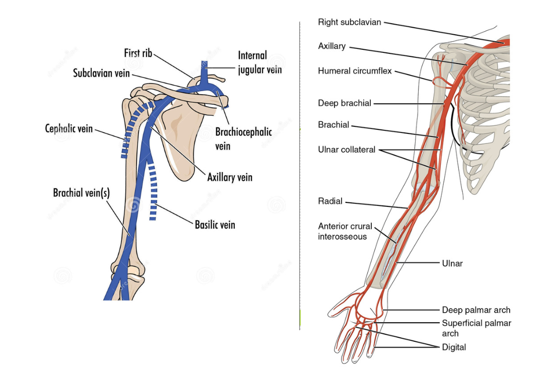
ARTERIES OF UPPER LIMB
Q.1 What is the extent of the axillary artery?
It extends from the outer border of the first rib to the lower border of teres major muscle. It is a continuation of the subclavian artery and it continues as a brachial artery.
Q.2 What are the parts of the axillary artery?
Pectoralis minor muscle crosses it and divides it into three parts:
Q.3 What are the relations of various nerves with the axillary artery?
First part:
Third part:
Q.4 What is the relation of various muscles with the axillary artery?
Anteriorly:
Posteriorly:
Laterally:
Q.5 Which veins cross the axillary artery?
Cephalic vein and thoracoacromial vein, the tributaries of axillary vein cross the first part.
Q.6 Name the branches of the axillary artery.
From first part:
From second part:
From third part:
Q.7 What is the extent of the brachial artery?
It extends from the lower border of teres major muscle to elbow at the level of the neck of radius just medial to the tendon of biceps.
Q.8 What are the nerves related to the brachial artery in its course?
Q.9 Name the branches of the brachial artery.
Q.10 What is the clinical importance of the brachial artery?
Brachial pulsations are auscultated in front of elbow just medial to the tendon of biceps while recording the blood pressure.
Q.11 Name the branches of profunda brachii artery.
Q.12 Name the branches of the radial artery.
Q.13 Where the pulsations of the radial artery are felt?
At the wrist against the anterior surface of the lower end of radius.
Q.14 Name the branches of the ulnar artery.
Q.15 Anterior interosseous artery lies between which muscles?
Flexor digitorum profundus and flexor pollicis longus.
Q.16 Name the structure separating the ulnar artery from the median nerve at the elbow.
Ulnar head of pronator teres.
Q.17 What are the relations of the radial artery in the forearm.
Radial artery lies between flexor carpi radialis and brachioradialis.
Q.18 What is the clinical importance of Allen’s test?
This test is performed to test the patency of the radial or ulnar artery.
Q.19 Name the arteries forming an anastomosis around the scapula.
Q.20 How the circulation is carried out when axillary artery is ligated.
By the collateral channels around the scapula, which connect the first part of the subclavian artery with the third part of the axillary artery.
VENOUS DRAINAGE OF UPPER LIMB
Q.1 What are the main superficial veins of the upper limb?
Cephalic (Preaxial) vein: Begins from the lateral end of the dorsal venous arch and drains into the axillary vein
Basilic (Postaxial) veins: Begins from the medial end of the dorsal venous arch and above the lower border of teres major continues as the axillary vein
Q.12 Name the structures lying between the axillary artery and vein.
The vein lies anteromedial to the artery.
The two are separated by:
Q.3 How the venous drainage of the upper limb is maintained in axillary vein obstruction?
By communication between the upper part of the cephalic vein with the external jugular vein in the neck.
Q.4 What is the clinical importance of the median cubital vein?
It is connected to the deep veins of the upper limb through a perforator which pierces the bicipital aponeurosis, which fixes it. So it does not slip away when intravenous injections are given.
LYMPHATIC DRAINAGE OF UPPER LIMB
Q.1 Which is the main lymph node of the upper limb?
Lateral group of axillary nodes.
Q.2 What is the area of lymphatic drainage of axillary lymph nodes?
Q.3 How the axillary lymph nodes drain?
Anterior, posterior and lateral group drain into the central group which in turn drains into the apical group. Subclavian lymphatic trunk from apical group drains into thoracic duct on the left side and to the right lymphatic duct on the right side.
Also read: Anatomy Question Collection
Also read: Anatomy Questions & Answers
Also read: Anatomy notes
Comments (0)