The Function of Spleen in Lymphatic System
5 years ago 109826
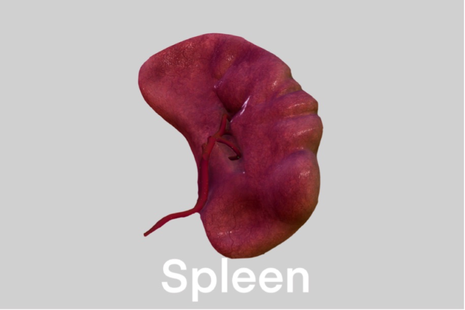
The spleen is a hemolymph organ and belongs to the reticuloendothelial system. And The Function of Spleen in Lymphatic System. It is situated in the upper and left part of the abdomen between the fundus of the stomach and the diaphragm.
During fetal life it produces erythrocyte & after birth, it produces T & B lymphocytes & belongs to the reticuloendothelial system.
| Two ends- Medial and Lateral |
| Two surfaces- Diaphragmatic and Visceral |
| Two borders- Superior and Inferior |
| Two angles- Anterior basal and posterior basal. |
| The diaphragm is separated by a recess of the greater sac of the peritoneum. |
| 9th, 10th, and 11th ribs of the left sides and their intercostal spaces. |
| In upper one-third - Lower border of the left lung and left pleural sac. |
| In lower two-third - Costo-diaphragmatic recess of left pleural sac |
4 impressions: Gastric, Renal, Colic, and Pancreatic.
| Fundus of the stomach in gastric impression. |
| Tail of pancreas in pancreatic impression. |
| Upper and lateral part of the anterior surface of the left kidney |
| Left colic flexure in the colic impression |
| Extension: From the hilum of the spleen to the greater curvature of the stomach. |
| Contents: short gastric, left gastro-epiploic vessels, associated lymphatics, and sympathetic nerves. |
2. Lieno-renal ligament -
| Extension: Extends from the hilum of the spleen to the anterior surface of the left kidney. |
| Contents: Splenic vessels, nerves, lymphatics. Sometimes the tail of the pancreas |
Suspends the spleen from above, known as the suspensory ligament of the spleen.
4. Phrenico-colic ligament:
It is not attached to the spleen, but indirectly supports the spleen from below, called sustentaculum lienis.
| 1. Open/Slow Circulation: |
| Central arteriole → Penicillar artery → Open space around red pulp → Venous sinusoid → Trabecular Vein → Splenic vein |
| 2. Closed/Fast Circulation: |
| Central arteriole → Penicillar artery → Venous Sinusoid → Trabecular vein → Splenic vein |
Identifying points
| Thick capsule and trabeculae present |
| Not differentiated into cortex and medulla. |
| Red pulp and white pulp are present. |
| White pulp (Malpighian body) has ecentric arteriole. |
Spleen is composed of Capsule & Parenchyma. Capsule extended down to form the trabeculae, which give it a lobular appearance.
Whereas Parenchyma has two components.
T & B lymphocytes aggregated around the central arterioles; (which branched from the trabecular artery) & form the White Pulp.
| 1. T-cells around the central arteriole. |
| 2. B-cells are arranged in the middle of the white pulp with the germinal center, where the germinal center is observed as a lightly stained area over the histological slide. |
| 3. Dendritic antigen-presenting cells around the periphery. |
On antigen stimulation, B-cells proliferate to form a germinal center where plasma cells also appear by differentiation.
RED PULP:
About 75% of the splenic parenchyma is formed by red pulp and contains large number of venous sinusoids.
The spaces between the venous sinuses are filled with an irregular and interconnected network of fibrocellular cords known as BILLROTH'S cords (Splenic Cord) consists of a labyrinth of macrophages.
| 1. Venous Sinusoids (Fenestrated porous capillary bulb) |
| 2. Splenic cord is mainly composed of reticular fibers & cells (which is the framework of lymphoid tissues), macrophages, Blood (i.e. RBC & other leukocytes). |
Red Pulp is like an open space cavity filled with blood.
In the Splenic cord, macrophages are present which phagocytose the damaged, expired, RBC(erythrocyte). So, the chief & important function of Spleen is to filter out the blood & scavenger of RBC(erythrocytes).
Artery supply:
Tortuous splenic artery (the largest branch of the coeliac trunk)
Other tortuous arteries of our body:
Venous drainage :
Drain into a splenic vein then into the trunk of the portal vein.
| 1. Filter blood by removing worn-out RBC & micro agents - (which occur around Red Pulp). |
| 2. Produce new lymphocytes which are later transported through blood-stream to target site - (Production occurs around White Pulp). |
Develops from the thickening of the mesenchyme in the dorsal mesentery.
Splenic notch, present in the upper border of the spleen which indicates that the spleen is lobulated in development.
Why during splenectomy damage to the tail of the pancreas have to be carefully avoided?
The presence of the tail of the pancreas in the splenicorenal ligaments sometimes results in its damage during splenectomy. The damaged pancreas releases enzymes that start to digest surrounding tissues with serious consequences like acute peritonitis.
Also read: Anatomy Question Collection
Also read: Anatomy Questions & Answers
Also read: Anatomy notes
Comments (0)