Embryology Notes From first week of development up to development of fetal membrane
6 years ago 5072
The first week of development
Ovarian cycle: At puberty, in every month ovary shows a cyclical change under the influence of gonadotropin-releasing hormone, from the hypothalamus. GRH acts on the anterior pituitary to release FSH and LH. The changes in the ovary every month known as the ovarian cycle.
FSH: follicle growth and maturation, secretion of estrogen, proliferative stage of menstruation.
LH causes ovulation, stimulate the production of progesterone.
Ovulation: shedding of the secondary oocyte (graffian follicle of ovum) with surrounding granulosa cells by rupturing the ovarian wall into the abdominal cavity is known as ovulation.
Time estimation :15+_or – 2 days
Cause: LH Surge
Clinically: ovulation can be felt by ovulation pain and increased body temperature 1 F
Corpus luteum:
After ovulation, granulosa cells remain in the wall of the ruptured follicle together with cells from theca interna, become vascularized and develop and a yellowish pigment called Corpus luteum.
Function: secrete progesterone and maintain the secretory stage of menstruation.
Types
Fertilization: It is the process by which mature male gamete fused with the mature female gamete to form a zygote in the ampullary region of the uterine tube. ( Because it is the widest part of the tube and closed to the ovary)
Prerequisite for fertilization
Phases of fertilization
Results of fertilization
Cleavage: It is the process by which one cell zygote undergoes a series of mitotic divisions.
Blastomere: The cells which become smaller with each cleavage division are known as blastomere.
Morula: 16 cell stage is known as a morula.
Blastocyst: When fluid begins to appear in the intercellular spaces of the inner cell mass, they confluent to form a single cavity, blastocele, at this time the embryo is called the blastocyst. The Zona pellucida disappears.
Blastocyst:
Inner cell mass (embryoblast)
Outer cell mass(trophoblast)??become the placenta
Implantation: it is the process by which blastocyst embedded on the mucus membrane of the uterine wall.
Site: It occurs on the anterior or posterior wall of the uterus near the fundus. On the 6th day of development.
Abnormal sites
Note: when implantation occurs other than normal site is known as abnormal implantation.
Ectopic pregnancy other than the uterus
Bilaminar germ disc: Epiblast and hypoblast together form bilaminar germ disc.
Decidua: Endometrium of the uterus at the time of pregnancy is known as decidua.
Decidual reaction: During pregnancy, cells of the endometrium become polyhedral, loaded with glycogen and lipid, intercellular space filled with extravasate and the tissue is redementus . These changes are known as a decidual reaction.
Events of 3rd week:
Gastrulation: The process that establishes all three germ layers (ectoderm, mesoderm, and endoderm) in the embryo is called gastrulation.
Process:
It begins with the formation of the premative streak on the surface of the epiblast. The premative streak is a narrow groove with a slightly raised area on either side. Cells of the epiblast migrate towards the premative streak and pass through it, this inward movement is known as invagination. Some invagination cells displace the hypoblast and create the embryonic endoderm. Cells come to lie between the epiblast and endoderm to form mesoderm. Cells in the epiblast become ectoderm.
Ectoderm-3 parts
Surface ectoderm: Epidermis of the skin including hairs, nails, sebaceous glands, sweat glands, lining epithelium of cheek, gum, mouth, nasal cavity, paranasal air sinuses, salivary glands, the lower part of the anal canal and urethra.
Neuro ectoderm
Ectodermal placid:
Lens placode-lens
Otic placode-internal ear
Neurulation: The conversion of the neural plate into a neural tube by folding is known as neurulation.
Process:
Due to the influence of notochord, overlying ectoderm thickened to form the neural plate. The lateral edges of the neural plate become more elevated to form neural folds and neural groove forms in the mid-region. Gradually neural folds fused and neural tube formed. Cephalic opening (24th day) of the neural tube is known as cranial neuropore. And caudal opening (27th day) is known as caudal neuropore. Finally the neuropore is closed.
The fate of the neural tube: The cranial portion becomes the brain and caudal becomes the spinal cord.
Ans. Exposure of brain tissue due to the absence of cranial vault. It is due to the failure of closure of cranial neuropore.
Spina – bifida: If caudal neuropore doesn’t close then spina bifida develops.
Neural crest: cells of the lateral border or crest of the neuroectoderm is known as neural crest.
Process of notochord formation:
Mesoderm:
Somite: segmental blocks of paraxial mesoderm are called somite. There are 42-44 pairs of somites.
Divided into three parts;
Each somite has its own sclerotome, myotome, and dermatome.
Derivatives of mesoderm
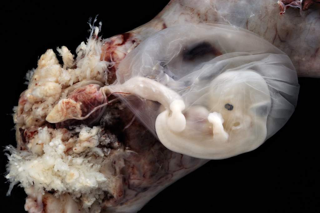
Fetal membrane:
Morphology of placenta:
Shape: discoid
Diameter:15-25cm
Thickness:3cm
Weight:500-600gm
Surface: fetal surface – smooth, covered by the amnion, and present the attachment of the umbilical cord.
Maternal surface: rough and irregular, present 15 to 25 cotyledons covered by decidua basalis.
Structure: chorionic plate on the fetal side, basal plate on the maternal side. Between them lies stem villi. Between the stem, villi lie maternal blood.
Chorionic plate:
Urachus: when the lumen of the allantois is obliterated a thick fibrous cord remains and connects the apex of the bladder with umbilicus is known as urachus.
Basal plate:
Development of placenta:
Maternal side- decidua basalis
Fetal side- chorionic frondosum
Decidua: Endometrium of the uterus at the time of pregnancy is called decidua.
Type: 3 types
Chorione: 1. Chorionic leabe- smooth
Note: human placenta – haemochorial type
Placental function:
Placental barrier:
Allantois: As a result of cephalocaudal and lateral folding of the embryo, a portion of the endoderm lined cavity remain outside the embryo known as the allantois.
Comments (0)