Basal Nuclei/ Basal Ganglia:
The basal nuclei are intra-cerebral masses of grey matter situated within the white core of the cerebral hemisphere which forms important parts of the extrapyramidal system.
Components:
1. Corpus striatum
- Caudate nucleus
- Lentiform nucleus
Larger lateral part - Putamen
Medial smaller part - Globus pallidus |
| 2. Amygdaloid body |
| 3. Claustrum |
Note:
| Functionally basal ganglia also include the substantia nigra of the midbrain & subthalamic nuclei of the diencephalon. |
| The Corpus striatum is divided into lentiform & caudate nuclei by a band of nerve fibers called an internal capsule. |
The function of Basal Nuclei:
| Control automatic & associated movements. (such as swinging of arms during walking) |
| Influences premotor cortex to control extrapyramidal activities |
| It controls abnormal involuntary movements |
| It controls group movement for emotional expression |
| Concerned with planning & programming of voluntary movement |
|
Inhibit unwanted muscular activity & decrease muscle tone
|
Note:
The basal ganglia do not receive any sensory input from the spinal cord.
Without sending fibers to the spinal cord modify the effect on the spinal cord through projection to the motor cortex & extra-pyramidal fibers.
They do not initiate movements but are able to adjust the motor commands.
* Basal ganglia are sometimes termed as extrapyramidal nuclei as it influences the quality of motor performance.
Connection of Basal Nuclei or Corpus Striatum with other parts
The caudate nucleus & putamen is the main afferent nuclei while globus pallidus is the outflow part (efferent part).
* Caudate nucleus receives fibers mainly from the cerebral cortex, thalamus & substantia nigra.
* Globus pallidus sends fibers to the thalamus, subthalamus, substantia nigra, and red nucleus.
| Different pathways are related to different neurotransmitters like dopamine, glutamate, acetylcholine & GABA. |
| Nigrostriate fibers are related to the dopamine pathway. |
Main Afferents Fibers:
Corticostriate Fibres:
The caudate nucleus & putamen receives axons from the cerebral cortex. |
Thalamostriate Fibers:
Thalamus sends a large no. of axons to the caudate nucleus & putamen. |
|
Nigrostriatal fibers:
Neurons in the substantia nigra send axons to the caudate nucleus & putamen.
|
Important efferent fibers:
Striatonigral fibers:
Fibers pass from the caudate nucleus & putamen to the substantia nigra |
|
Straiatopallidal fibers:
Fibers from caudate & putamen to globus pallidus.
|
Disorders of basal nuclei:
Lesions of the basal ganglia & cerebellum do not cause paralysis.
* These produce abnormal movements or unwanted involuntary movements or posture or change in tone.
Hyperkinetic disorders:
Excessive & abnormal movements like chorea, athetosis, ballism
Hypokinetic disorder:
There is a lack of slowness of movements such as Parkinson’s disease.
Parkinson’s disease:
Occur due to Dopamine deficiency in the corpus striatum resulting from the destruction of substantia nigra or lesion in nigrostriatal fibers.
Sign-symptoms
- Hypertonicity or muscular rigidity
- Involuntary tremors / Resting tremors
- Gait disturbance
- Akinesia:
Voluntary movements may be impossible.
Serious difficulty in initiating movements
- Loss of facial expression / Mask like face
L-dopa (a precursor of dopamine) is used as a replacement therapy for Parkinsonism.
Because dopamine, the normal neurotransmitter in the striatum, is reduced in these cases.
The nigrostriatal fibers are considered important in the genesis of Parkinsonism tremor since its neurons utilize dopamine in the neurotransmission.
| Dopamine is not directly used in treatment because of its high polarity, it cannot easily cross the Blood-Brain Barrier (BBB). |
| It is very hard to treat the patient of the brain because normal drugs cannot cross the BBB easily. |
Chorea:
This means dancing involuntary movements
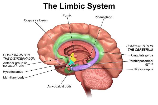
Limbic system:
The limbic system means the entire neural circulation that controls the emotional, behavioral & motivational drives.
(Limbus=means ring-shaped)
The limbic lobe is a composite bordering zone between the cerebrum and diencephalon.
* It is somehow ring-shaped.
* It is associated with a basic survival behavioral control system
Constituents of the limbic system:
| Olfactory nerve, Olfactory bulb & tract |
| Septal region |
| The anterior nucleus of the thalamus |
| Mammilothalamic tract |
| Hypothalamus (mainly mammillary bodies) |
| Limbic Lobe consists of the cingulate gyrus, uncus, parahippocampal gyrus |
| Hippocampus Formation including the hippocampus, dentate gyrus, longitudinal striae, indusium griseum |
| Fornix |
Tips:
The mammilothalamic tract is the bundle of nerve fibers connecting the mammillary body & anterior nucleus of the thalamus.
The function of the limbic system
The main object of the limbic system is to meet the needs of primitive life that is food and sex.
Food is necessary for the survival of individuals & sex for the survival of the species.
- It controls food habits necessary for the survival of the individual.
- It controls the sex behavior necessary for the survival of the species.
- It controls emotional behavior like joy, sorrow, and fear.
- Convert recent memory to long-term memory.
Applied anatomy:
| The hippocampus can be regarded as the cortical center for autonomic reflexes. |
|
The hippocampal amygdala complex is related to the memory of recent events. Lesions related to a loss of recent memory only.
The patient is unable to commit any new facts to memory and does not remember recent events.
|
|
Destruction of the olfactory nerve results in loss of the sense of smell (anosmia).
|
Interpeduncular fossa:
The interpeduncular fossa is a rhomboid-shaped fossa when seen from the inferior surface of the base of the brain.
Boundary:
|
Anteriorly:
Optic chiasma
|
|
Anterolaterally:
Optic tract
|
|
Posterolaterally:
Crus cerebri of midbrain
|
|
Posteriorly:
The upper part of Pons
|
Content of fossa
- Mammillary body
- Posterior perforated substances
- Infundibulum
(A narrow stalk that connects the pituitary gland)
- Oculomotor nerve
Note: This fossa is surrounded by a circle of Willis
Clinical importance:
In head injury, the contents of the interpeduncular fossa is usually injured.
Comments (0)