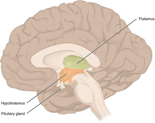
Diencephalon (Thalamus & Hypothalamus)
Part of Forebrain
Diencephalon:
The cavity within the diencephalon - 3rd ventricle.
* This 3rd ventricle makes the division of the diencephalon into right and left symmetrical parts.
The hypothalamic sulcus that extends from the interventricular foramen to the cerebral aqueduct makes a division of each side of the diencephalon into
The dorsal part of the Diencephalon
- Thalamus
- Metathalamus
(Medial & Lateral geniculate bodies)
- Epithalamus, including the pineal body & habenula
The ventral part of the diencephalon
Thalamus:
Thalamus - a huge mass of grey matter.
Consists of multiple nuclei.
* It forms the lateral wall of the third ventricle & also the floor of the central part of the lateral ventricle.
* On either side of the 3rd ventricle (a slit-like cavity), two thalami are present.
* On the posterior part, it overhangs the lateral and medial geniculate bodies and the superior colliculus with its brachium.
Surface:
Medial Surface:
Form Maximum part of the lateral wall of the 3rd ventricle.
Lateral Surface:
Forms the medial boundary of the internal capsule.
Note:
| Interthalamic adhesion - Interconnect the medial surface of both thalami. |
|
Thamalus forms the lateral wall of the 3rd ventricle in the posterosuperior part.
At the same time, anteroinferior, the hypothalamus forms the lateral wall of the 3rd ventricle.
|
Nuclei of the thalamus:
| Anterior part |
Anterior nucleus |
| Medial Part |
Medial Ventral nucleus
Medial dorsal nucleus |
| Lateral part |
------------- |
| *Dorsal part |
Lateral dorsal
Lateral posterior
Pulvinar |
| *Ventral part |
Ventral anterior
Ventral lateral
Ventral posterolateral
Ventral posteromedial |
The ventral posteromedial nucleus receives:
- General sensory impulses from the face and head through the trigeminal lemniscus
- Tastes sensations from taste buds through solitariothalamic tract
The ventral posterolateral nucleus receives:
- Exteroceptive sensations (pain, touch, temperature) through spinal lemniscus
- Proprioceptive sensations through medial lemniscus. (ie. muscle and joint sense, vibration, two-point discrimination)
(from the rest of the body except the face and head)
Important Connection of Thalamus:
| Ventral Posterolateral nucleus |
Trigeminal lemniscus
Solitariothalamic lemniscus
|
To postcentral gyrus (area no. 3,1,2) |
Relay station for impulses from face & taste impulse |
| Ventral Posteromedial Nucleus |
Spinal lemniscus
Medial lemniscus |
To postcentral gyrus (area no. 3,1,2) |
Relay station for exteroceptive & proprioceptive impulses from body, except face
|
Thus, the thalamus is considered as the chief integrating center where sensory impulses from all these sources is brought together.
* All sensory impulses reaching the ventral posterior nucleus complex are carried to the primary sensory area of the cerebral cortex by fibers passing through the posterior limb of the internal capsule.
The function of the Thalamus as a whole:
It acts as a sensory relay station for all the sensory pathways except for the olfactory pathway.
Thalamus is considered as the great sensory gateway to the cerebral cortex.
It receives sensory impulses from the opposite half of the body and transmits most of them to the Somatosensory area of the cerebral cortex (Brodmann area no.3, 2,1).
|
Note:
4 lemniscus system in the brain stem:
Medial Lemniscus
Spinal Lemniscus
Lateral Lemniscus
Trigeminal lemniscus |
|
Medial lemniscus comprises of Tract of Gall & Burdach.
|
| Spinal Lemniscus comprises of Anterior & Lateral spinothalamic tract. |
| The major sensory supply in the face is by the three divisions of the trigeminal nucleus. |
Exteroceptive Sensation - (Pain, Temperature, Pressure, Touch)
|
| Proprioceptive Sensation - (Muscle & joint sense, Vibration, Two-point discrimination) |
Metathalamus:
Consists of medial & lateral geniculate bodies present on either side of the midbrain, below the thalamus.
Medial geniculate body:
Present medial to the midbrain
* Medial geniculate body is connected to the inferior colliculus by the Inferior Brachium.
Connection:
Afferents
Lateral Lemniscus
Fibers from Inferior colliculus |
|
Efferents
Gives rise to auditory radiation
I
Sublentiform part of the internal capsule
I
Auditory area of the temporal lobe
|
Lateral Geniculate Body:
- Present anterolateral to the medial geniculate body, below the thalamus.
- The superior colliculus is connected to it by the superior brachium.
- Forms part of the visual pathway, the site of termination of the optic tract.
Connection:
Afferent:
Optic tract |
Efferent:
Give rise to optic radiation
I
Retrolentiform part of the internal capsule
I
The visual area of cortex |
Structure of Lateral geniculate:
It is a six-layer.
* Layers 1, 4 & 6 receive optic fibers from the opposite eye through the ipsilateral optic tract.
* Layers 2, 3 & 5 receive optic fibers from of the same side-eye through the ipsilateral optic tract
Note:
|
Nasal fibers cross at the optic chiasma and go to opposite sides.
|
|
Thus, the optic tract contains temporal fibers on the same sides and nasal fibers on the opposite side.
|
Function:
The lateral geniculate body is the last relay station of the visual pathway to the occipital cortex.
Summary of Pathway:
Auditory sense - Lateral lemniscus - Inferior colliculus - Medial geniculate body - Auditory radiation - Sublentiform part of the internal capsule - the auditory cortex
Visual sense - optic chiasma - optic tract - Lateral geniculate body - optic radiation - retro-lentiform part of the internal capsule - visual cortex
| Part |
Afferent Fiber |
Efferent fiber |
Function |
| Medial geniculate body |
Auditory fibers from the inferior colliculus |
Primary auditory area (41,42) |
Relay station for auditory impulses |
| Lateral geniculate body |
Optic tract |
Primary Visual Area (17) |
Relay station for visual impulses |
Corpora quadrigemina:
In the brain, four colliculi (two superior & two inferior) are located on the tectum at the dorsal aspect of the midbrain called Corpora Quadrigemina.
- Superior colliculus related to visual reflexes
- Inferior colliculus form an auditory pathway
* Superior colliculus
I
superior brachium - medial geniculate body
* Inferior colliculus -
I
inferior brachium - lateral geniculate body
Epithalamus:
Part:
- Right & left habenular nuclei
- Pineal body
- Habenular commissure
- Posterior commissure
Pineal body /Pineal Gland:
The pineal gland is the small body projecting backward and downwards between the two superior colliculi.
* Pineal gland is the neuroendocrine gland that contains parenchymal cells called pinealocytes and neuroglia cells.
* It is the only part of the brain without neural tissue in it.
The function of pineal gland:
- Pineal gland secretion including melatonin has inhibitory influences on endocrine glands and gonads.
- It secretes hormones called melatonin which cause changes in skin color in some species.
Hypothalamus:
The hypothalamus is a part of the diencephalon.
It lies in the floor and lateral wall of the third ventricle.
Anatomically, it includes:
a. The floor of the third ventricle
b. The lateral wall of the third ventricle below the hypothalamic sulcus.
|
It means:
The floor and lateral wall of the 3rd ventricle are also part of the hypothalamus.
|
Boundaries
* Superiorly:
Thalamus
* Inferiorly:
The floor of 3rd ventricle
* Posteriosuperiorly:
Hypothalamic sulcus
* Anteriorly:
Lamina terminalis (extends from otic chiasma to anterior commissure)
Parts & Nuclei of Hypothalamus:
| Optic part |
1. Preoptic nucleus
2. Supraoptic nucleus
3. Paraventricular nucleus
4. Anterior nucleus |
| Mammillary part |
1. Arcuate nucleus
2. Ventromedial nucleus
3. Dorsomedial nucleus |
| Tuberal part |
1. Posterior nucleus
2. Mammillary nuclei
3. Lateral nucleus |
Connection of hypothalamus:
Axons of para-ventricular nuclei & Supraoptic nuclei go through the pituitary stalk forming the hypothalami-hypophyseal tract & go downwards to the posterior pituitary gland (neurohypophysis).
* Vasopressin & oxytocin (synthesized in supraoptic & paraventricular nuclei) are transported through this tract to the posterior pituitary gland.
Axons of other nuclei transfer their secretion to the hypothalamohypophyseal portal system of blood vessels.
* And controls hormone secretion from the anterior pituitary gland (adenohypophysis).
Note:
|
Vasopressin & oxytocin are the hormones secreted from the hypothalamus, but they remain stored in the nerve fibers ending in the hypothalami-hypophyseal tract & this tract descends up to the posterior pituitary gland.
* Thus, in a simple manner, it can be said that the posterior pituitary gland acts as a storehouse for vasopressin(ADH) & oxytocin.
|
|
When nerve fibers get stimulated, then these tracts drain hormones to the systemic circulation.
|
|
Tracts are the bundle of nerve fibers (axons or dendrites) within the CNS.
|
Function of the hypothalamus:
1) Autonomic control
- The anterior part controls the parasympathetic nervous system.
- The posterior part of the hypothalamus controls the sympathetic nervous system.
Thus, the hypothalamus controls the cardiovascular, respiratory, and alimentary functions.
2) Regulation of body temperature
- The anterior part helps in decreasing body temperature by causing cutaneous vasodilation & increases sweating
The posterior part of the hypothalamus is stimulated in cold climates increasing sympathetic activity.
|
Causes vasoconstriction of skin blood vessels
|
Decreases heat loss & sweating
3) Regulation of sensation of thirst & water intake
- The lateral part of the hypothalamus acts as a thirst center
4) Regulation of Hunger/Food intake
- The feeding center is placed laterally. Stimulation of the feeding center causes hyperphagia (overeating)
- The satiety center is placed medially, stimulation of this causes hypophagia.
5) Regulation of endocrine control
- By secreting releasing & inhibiting hormones, it controls the secretion from the anterior pituitary gland.
- Formation of posterior pituitary gland hormone & their regulation of secretion (neurosecretion).
6) Reproduction & sexual behavior:
- Regulates the secretion of gonadotrophin hormones by the pituitary gland influencing sexual behavior & reproduction
7) Biological clock:
- Cyclic activity regulation is like the sleeping & waking cycle.
Simply,
1) Regulation of Endocrine Function by secreting releasing & inhibiting hormones.
2) Regulation of body temperature
3) Regulation of the Cardiovascular system
4) Regulation of water intake & sensation of thirst
5) Regulation of hunger, feeding & GI activity
The lesion in hypothalamus could cause
1) Obesity
2) Hyperglycemia & glycosuria
3) Diabetes insipidus
4) Sexual disturbance
5) Sleep disturbances
Comments (0)