Orbit and eye muscles
Orbit:
Boundaries of orbit
Roof: By
- Orbital plate of the frontal bone
- Lesser wing of sphenoid bone
Medial wall: By
- Maxilla
- Lacrimal bone
- Ethmoid bone
- Body of sphenoid
Floor: By
- Orbital surface of the zygomatic bone
- Orbital surface of the maxillary bone
- Orbital process of palatine bone
Lateral wall: By
- Zygomatic bone
- Greater wing of the sphenoid
Feature present in Boundary wall:
On medial wall:
The lacrimal fossa is present between the frontal process of the maxilla and the lacrimal bone.
It lodges for the lacrimal sac.
The nasolacrimal canal lodges for the nasolacrimal duct which opens in the inferior nasal meatus of the nasal cavity.
On the floor:
Infraorbital groove and canal which contain infraorbital nerve and vessels, branches of maxillary nerve and vessels.
On the roof:
- Fossa for lacrimal gland in the anterolateral part
- Trochlear notch at the anteromedian part
- Optic canal lies at the most posterior part of the roof
(Apex of orbit lies between optic canal & superior orbital fissure)
Content of orbit:
1) Eyeball (most important content)
2) Fascia (orbital & bulbar)
3) Muscles:
Extraocular & Intraocular
4) Vessels:
Ophthalmic artery
Superior & inferior ophthalmic vein
5) Nerves:
Optic, Trochlear, Abducent nerve
Branches of the ophthalmic nerve, &
Sympathetic nerves
Ciliary ganglia
6) Lacrimal gland
7) Orbital Fat
Note:
| Bulbar fascia also called tenon’s capsule means fascia around the bulb of the eyeball, cover the posterior outer surface of the sclera which cannot be seen. |
| The outer anterior surface of the sclera which can be seen through the eye is covered by conjunctiva. |
Foramen opening in orbit:
Structure passing through optic canal
- Optic nerve
- Ophthalmic artery, 1st branch of internal carotid artery
Structure passing through superior orbital fissure
- Oculomotor nerve
- Trochlear nerve
- Abducent nerve
- Branches of ophthalmic nerve
1. Frontal nerve
2. Lacrimal nerve
3. Nasociliary nerve
- Superior Ophthalmic vein, which drains to the cavernous sinus
Structure passing through inferior orbital fissure
- Infra-orbital nerve and zygomatic nerve, branches of maxillary division of Trigeminal nerve
- Infra-orbital artery and vein, branches of the maxillary artery of external carotid artery
-
Inferior ophthalmic vein
Note:
| Inferior orbital fissure doesn’t open into the cranial fossa. Thus it cannot be seen from the superior view of the cranial fossa. |
|
Both the optic canal and superior orbital fissure communicate into middle cranial fossa.
|
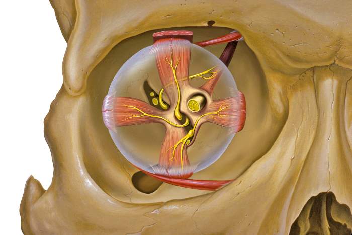
TYPES OF EXTRAOCULAR MUSCLES:
Voluntary Muscles
1) Four recti:
-
Superior rectus
-
Inferior rectus
-
Medial rectus
-
Lateral rectus
2) Two oblique:
- Superior oblique
- Interior oblique
3) The levator palpebrae superioris elevates the upper eyelid.
Involuntary Muscles
- Superior tarsal muscle - elevate upper eyelid
- Inferior tarsal muscle - depresses the lower eyelid
- Orbitalis
Note:
|
Levator palpebrae superioris is the muscle of facial expression.
|
|
All the muscles of facial expression are supplied by facial nerve except levator palpebrae superioris which is supplied by the oculomotor nerve.
|
| All recti muscles arise from the corresponding margins of the common tendinous ring (of Zinn). |
|
All recti muscles inserted just posterior to the junction between sclera and cornea (corneoscleral junction also called as limbus).
|
Nerve supply of Extraocular muscles:
All the Extraocular muscles are supplied by oculomotor nerve except superior oblique (by Trochlear nerve) & lateral rectus (by Abducent nerve)
Mnemonic: All are 3 except SO4, LR6
Intraocular muscle
| Intraocular Muscle |
Nerve Innervation |
Function |
| Ciliary muscle |
Oculomotor (parasympathetic) through ciliary ganglion |
Control shapes of lens
Accommodation
|
| Sphincter pupillae of iris |
Oculomotor (parasympathetic) through ciliary ganglion |
Constricts pupil |
| Dilator pupillae of iris |
Cervical sympathetic fibers that have comes along the internal carotid artery |
Dilates the pupil |
Movement of eye:
a. Upward rotation or elevation:
By the superior rectus and the inferior oblique
b. Downward rotation or depression:
By the inferior rectus and the superior oblique
c. Medial rotation or adduction:
By the medial rectus, the superior rectus and the inferior rectus
d. Lateral rotation or abduction:
By the lateral rectus, the superior oblique, and the inferior oblique
e. Intortion:
By the superior oblique and the superior rectus
f. Extortion:
By the inferior oblique and the inferior rectus.
Action of Extraocular muscles
| Muscles |
Movements |
| Superior rectus: |
Elevation, medial rotation, Intorsion |
|
Inferior rectus:
|
Depression, medial rotation, Extorsion
|
| Inferior oblique: |
Elevate, lateral rotation, Extorsion |
| Superior oblique: |
Depression, Lateral rotation, Intorsion |
| Medial rectus: |
medial rotation |
| Lateral rectus: |
lateral rotation |
Notes:
Torsion is the rotatory movement around the anteroposterior axis.
When 12 o'clock position of the cornea rotates medially, it is intorsion.
When 12 o'clock position of the cornea rotates laterally, it is extorsion. |
|
Medial and lateral rotation occurs in the vertical axis but intortion and extortion (a type of rotation) occur in the anteroposterior axis.
|
| Conjugate movement: When both eyes move in the same direction with the visual axis being parallel. |
Vessels of orbit:
Ophthalmic artery
- Branch of internal carotid artery
- Passes through optic canal along with optic nerve
Major branches of the ophthalmic artery:
- Central artery of retina
- Posterior(long & short) ciliary arteries which chiefly supplies the choroid and iris
- Supraorbital & Supratrochlear which passes through supraorbital notch and supratrochlear notch respectively.
- Central artery of retina
The central artery of the retina pierces the dural sheath of the optic nerve and enters into the substance of the nerve.
After reaching the retina, it divided into branches that supply the retina.
The central artery of retina - an end artery
And it is the major blood supply to the retina. So, its occlusion can lead to blindness.
Ophthalmic vein:
Superior ophthalmic vein
|
Passes through superior orbital fissure
|
Drain into cavernous sinus
Inferior ophthalmic vein joins the superior ophthalmic vein and drain to the cavernous sinus
Or
Inferior ophthalmic vein
|
Passes through inferior orbital fissure
And join pterygoid plexus of vein
Nerves of Orbit:
- Optic
- Oculomotor with ciliary ganglion
- Trochlear
- Branches of ophthalmic and maxillary divisions of the trigeminal nerve
- Abducent nerve and
- Sympathetic nerves
Optic nerve:
Purely sensory
It is made up of the axons of cells in the ganglionic layer of the retina.
It passes through the optic canal to enter the middle cranial fossa where it joins the optic chiasma.
| The optic nerve is a tract, not a nerve in a strict sense. |
| It has no neurolemmal sheath and has no power of regeneration. |
| Developmentally, the optic nerve and the retina are a direct prolongation of the brain. |
| Whole nerves are coved by three meningeal sheath with subarachnoid space extending up to the eyeball. |
| The fibers of the optic nerve are myelinated by oligodendrocytes rather than Schwann cells. |
| Thus, the optic nerve is not a true peripheral nerve. |
Ciliary ganglion:
One of the peripheral parasympathetic ganglion among the four parasympathetic ganglion.
Lie near the apex of the orbit between the optic nerve and lateral rectus muscle.
Summary Connection:
Parasympathetic fibers arise from Edinger-Westphal nucleus
|
Transmit fibers to oculomotor nerve
|Relay on
|Ciliary ganglion
|
8-10 branches of short ciliary nerve arises from ganglion
|
Supply ciliaris muscle and sphincter pupillae of iris
|
Help in accommodation
Branches of ophthalmic division of Trigeminal nerve
Frontal nerve
- Supratrochlear
- Supraorbital
Nasociliary nerve
- Infratrochlear
- Posterior ethmoidal
- Anterior ethmoidal
- Sensory Branch to ciliary ganglion
- Long ciliary nerve
Lacrimal nerve
Pterygopalatine ganglion --Zygomatic nerve passes secretomotor fibers to the lacrimal nerve to supply the lacrimal gland.
Comments (0)