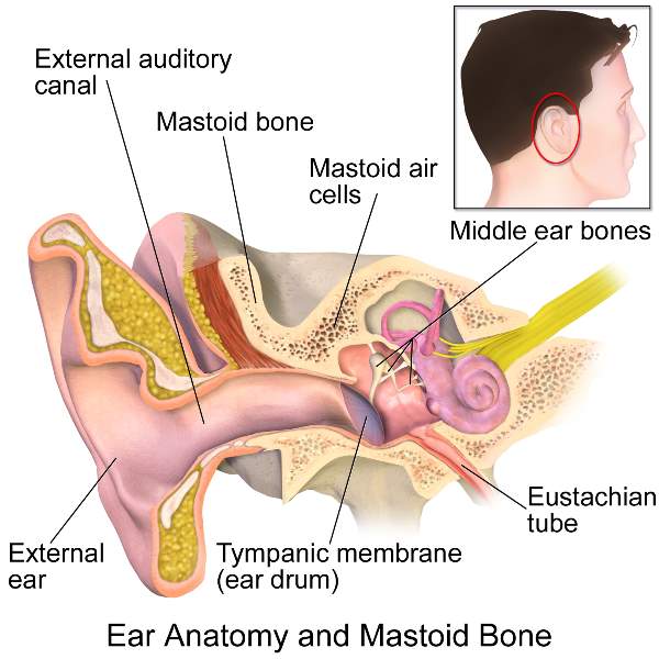
External and middle ear
Ear
The external ear consists of:
- Auricle/Pinna
- External acoustic meatus
Auricle
Made from a single plate of elastic cartilage.
However lowest part of the auricle is soft and consists only of fibrocartilagenous tissue and called the lobule for wearing the earrings.
Parts of auricle
External Acoustic Meatus
It is the channel for sound waves to reach the tympanic membrane.
It is about 24mm long in adult which is divided into the outer Cartilaginous part and inner bony part.
Cartilaginous part:
- Form outer 2/3 rd of the external auditory meatus
- 8mm in length
- Formed by elastic cartilage which is the continuation of the pinna.
Bony part:
- Formed by the tympanic plate & squamous part of the temporal bone.
- 16mm in length
[External acoustic meatus is lined by skin ie keratinized stratified squamous epithelium, which further continued to form the outer cuticular layer of the tympanic membrane]
Tympanic membrane (Eardrum)
- It is the thin, semi-transparent membrane that make partition between the external acoustic meatus and the middle ear.
- It is placed obliquely at an angle of 55 degrees with the floor of the meatus.
- Internally, the tympanic membrane is crossed by the chorda tympani nerve, branch of the facial nerve.
Direction:
Downwards, forwards and laterally
Attachment:
Its circumference is attached to the tympanic sulcus present in the petrous part of the temporal bone, but superiorly where sulcus is deficient is attached to tympanic notch.
From the ends of the notch, anterior and posterior malleolar folds arise.
- The greater part of the tympanic membrane is tightly stretched called pars tensa.
- The part between the two malleolar folds is loose and is called pars flaccid.
The membrane is held tense by the inward pull of the tensor tympani muscle which is inserted into the upper end of the handle of the malleus.
Surfaces
Outer surface lined by the thin skin.
The inner surface provides attachment to the handle of the malleus which extends up to its center.
[The point of maximum convexity lies at the tip of the handle of the malleus and is called umbo.]
Structure of Tympanic membrane
- Outer cuticular layer of skin
- Middle fibrous layer made up of superficial radiating fibers and deep circular fibers.
- Inner mucous layer lined by ciliated columnar epithelium
Lining epithelium:
1) External acoustic meatus:
Skin- Stratified squamous epithelium
2) Tympanic membrane
- Outer cuticular layer - Stratified squamous epithelium
- Inner mucous layer - Ciliated columnar epithelium
- Middle ear cavity - Ciliated columnar epithelium
3) Auditory tube - Ciliated columnar epithelium
Note:
As the outer cuticular layer is a continuation of skin that lined external auditory meatus, it is lined by keratinized stratified squamous epithelium.
The main component of the middle ear is filled by air.
Thus inner layer of the tympanic membrane is lined by ciliated columnar epithelium.
Function of tympanic membrane
It makes vibration of air or sound then transmit to the handle of the malleus which is attached to the eardrum and to the incus and stapes (ear ossicle).
Middle ear (Tympanic cavity)
Cavity of middle ear divided into:
- An epitympanic recess which lies above the level of the tympanic membrane
- Tympanic cavity proper which lie opposite to the tympanic membrane
Communication of middle ear cavity
- Anteriorly:
With nasopharynx through auditory tube
- Posteriorly:
With mastoid antrum and mastoid air cells through aditus (opening) to the mastoid antrum.
[Note: Auditory tube open on the lateral wall of the nasopharynx.]
Contents of middle ear:
- Ear ossicle/ bone
Malleus, Incus & stapes
- Ligaments of the ear ossicles
- Two muscles
Tensor tympani and the stapedius
- Vessels supplying and draining the middle ear
- Nerves
Chorda tympani nerve & Tympanic plexus
- Air
[Note: Ligament of ear ossicles need to stabilize the ossicles]
Boundaries of Middle ear cavity
Roof
- Formed by a thin plate of bone called as tegmen tympani of petrous part of temporal bone
- The roof separates the middle ear from the middle cranial fossa.
Note:
Tegmen tympani prolonged to form the roof of the canal of tensor tympani.
Tegmen tympani is elevated due to the underlying semicircular canal of the inner ear.
Floor / Jugular wall
- Formed by a thin plate of bone of petrous part of temporal bone
- Separate the middle ear from the superior bulb of the internal jugular vein.
Note:
Superior petrosal sinus drain into the transverse sinus
Inferior petrosal sinus drains into the superior bulb of the internal jugular vein
Anterior wall/ Carotid wall
- Uppermost part of the anterior wall bears the opening of the canal for the tensor tympani
- Middle part has an opening of the auditory tube
- The inferior part is formed by a thin plate of bone that forms the posterior wall of the carotid canal.
- This plate separates the middle ear from the internal carotid artery.
[Note: Anterior wall is directed toward the apex of the petrous part of the temporal bone.]
Posterior wall/ Mastoid wall
- An aditus to mastoid antrum
(An opening or aditus through which the middle ear cavity communicates with the mastoid antrum.)
Lateral wall / Membranous wall
- Mainly formed by tympanic membrane and partly by the squamous part of the temporal bone
- The lateral wall separates the middle ear from the external ear.
Medial wall
- Promontory
A rounded bulging produced by the first turn of the cochlea.
It is grooved by Tympanic plexus.
- Fenestra vestibuli (Oval window)
An oval opening which leads into the vestibule of the internal ear and is closed by the foot-plate of the stapes.
- Prominence of facial canal
It ultimately ends at the stylomastoid foramen.
- Fenestra cochleae (Round window)
A round opening which opens into the scala tympani of the cochlea and is closed by the secondary tympanic membrane.
- Medial wall separates the middle ear from the internal ear
Note:
|
Motor fibers of facial nerve exist through the stylomastoid foramen and supply the muscles of facial expression.
|
Ear ossicles:
Malleus, incus and stapes are bone within the bone, as these three bony ossicles lie within the petrous part of the temporal bone.
Malleus (hammer-shaped)
The handle of malleus is attached up to the upper half of the tympanic membrane
Stapes:
The footplate fits into the fenestra vestibuli (round window)
- This footplate act as the hydraulic pump piston which helps in transmitting the ear ossicles vibration into the perilymph of the scala vestibule.
[Mnemonic: MISs]
Synovial Joint of Ossicles
- Incudomalleolar joint (Saddle type of synovial joint)
- Incudostapedial joint (Ball and socket type of synovial joint)
Muscles of the Middle Ear
Tensor tympani
It lies in the canal for tensor tympani which lie above the auditory tube which forms the anterior wall of the middle ear
--Inserted to the handle of malleus
Stapedius
Inserted into the stapes bone
|
Tensor tympani muscle develops from 1st pharyngeal arch (mesodermal in origin) and is supplied by the mandibular division of the Trigeminal nerve.
|
|
Stapedius develops from the 2nd pharyngeal arch (mesodermal in origin) and is supplied by the facial nerve.
|
Note:
Structure derived from
Pharyngeal arches are mesodermal in origin.
Pharyngeal cleft (ectodermal)
Pharyngeal pouches (endodermal)
Function:
Both muscles act to damp down the intensity of high-pitched sound waves and thus protect the internal ear.
Clinical anatomy:
|
Hyperacusis:
Due to paralysis of the stapedius muscle, the stability of stapes is not maintained; so even small sounds get too high in volume.
|
Function of middle ear
It transmits sound waves from the external ear to the internal ear through the chain of ear ossicles, and thus transforms the air-borne vibrations from the tympanic membrane to liquid borne vibrations in the internal ear.
The intensity of the sound waves is increased ten times by the ossicles.
Tympanic or Mastoid Antrum
Mastoid antrum is a small, circular, air-filled space situated in the posterior part of the petrous temporal bone.
- It communicates anteriorly with to middle ear through aditus(opening) to mastoid antrum.
- The floor of antrum receives the opening of the mastoid air cells
Mastoid Air Cells
Mastoid air cells are a series of intercommunicating spaces of variable size present within the mastoid process.
Mastoid air cells communicate superior with mastoid antrum.
Comments (0)