Nose & Paranasal Sinuses
Nose
Lining epithelium in the nose
Upper 1/3rd of the cavity including the roof formed by cribriform palate
- Lined by olfactory mucosa which contains receptors called olfactory cells for the sense of smell
Rest of the nasal cavity
- Lined by respiratory mucosa (Pseudo-stratified ciliated columnar epithelium)
Lining epithelium of paranasal sinuses
Pseudo-stratified ciliated columnar epithelium
External Nose Formation:
1) Bony part (Forming upper part)
- Nasal bone
- Frontal process of maxilla
2) Cartilaginous part (Forming lower part)
- Superior nasal cartilage
- Inferior nasal cartilage
- Septal cartilage
- Alar cartilages
Nasal cavity
Extension
From external nasal nares/nostril to posterior nasal apertures (after which it is continued as nasopharynx)
Roof formation of Nasal Cavity
- Anterior slope
By nasal part of the frontal bone, nasal bone, and nasal cartilage
- Middle horizontal part
By cribriform plate of ethmoid
- Posterior slope
By the inferior part of the body of the sphenoid bone
Floor formation of Nasal cavity
- Anterior 2/3rd
By palatine process of maxilla
- Posterior 1/3rd
By horizontal plate of the palatine bone.
Nasal Septum Of Nasal Cavity
The nasal septum is the median osseocartilaginous partition making the two halves division of the nasal cavity.
Bony part is formed by
- Perpendicular plate of ethmoid bone
- Vomer
Cartilaginous part is formed by
- Septal cartilage
- Septal process of inferior nasal cartilage
Cuticular part is formed by fibrofatty tissue covered by skin.
Lateral wall of the Nasal Cavity
The lateral wall of the nose is irregular due to the presence of 3 shelf-like bony projections called conchae.
The conchae increasing the surface area of the nose makes the effectiveness of air-conditioning of the inspired air.
Lateral wall formation
1) Bony Part is formed by:
- Nasal bone
- Frontal process of the maxilla
- Lacrimal bone
- Labyrinth of ethmoid with superior and middle conchae
- Inferior nasal concha
- Perpendicular plate of palatine bone
- Medial pterygoid plate
2) Cartilaginous part is formed by:
- Superior nasal cartilage
- Inferior nasal cartilage
- 3/4 small alar cartilage
3) Cuticular part is formed by fibrofatty tissue covered with skin
Space present on lateral wall of nasal cavity
- Inferior nasal meatus
- Middle nasal meatus
- Superior nasal meatus
- Sphenoethmodial recess
Note:
[Each meatus communicates freely with the nasal cavity proper.]
Nasal Conchae:
Nasal conchae are curved shelf-like bony projections present on the lateral wall of the nasal cavity.
Superior nasal conchae and middle nasal conchae are the bony projections from the medial surface of the ethmoid labyrinth.
Inferior nasal conchae is a separate independent bone.
Nasal Meatus:
Meatuses of the nose are the space beneath the overhanging conchae.
[Or The space separating the conchae is called meatus]
Inferior nasal meatus
The space present in the lateral wall of the nasal cavity beneath the inferior nasal conchae is called inferior nasal conchae.
- Nasolacrimal duct opens in the junction of anterior 1/3 and posterior 2/3rd of the inferior meatus.
Middle nasal meatus
The space present in the lateral wall of the nasal cavity between the middle nasal conchae & the inferior nasal conchae is called the middle nasal meatus.
Opening present in middle nasal meatus:
- Opening of frontal air sinus
- Opening of maxillary sinus
- Opening of anterior ethmoid sinus
Superior nasal meatus:
The space present in the lateral wall of the nasal cavity between the superior nasal conchae & the middle nasal conchae is called the superior nasal meatus.
- Opening of Posterior ethmoid air sinus lies here.
Sphenoethmodial recess:
It is a triangular fossa just above the superior concha.
Here, Sphenoidal air sinus opens.
Structures opening into meatus or lateral wall of the nasal cavity
| Inferior nasal meatus |
Nasolacrimal duct |
| Middle nasal meatus |
Frontal air sinus
Maxillary air sinus
Anterior and middle ethmoidal sinus |
| Superior nasal meatus |
Posterior ethmoidal sinus |
| Sphenoethmodial recess |
Sphenoidal air sinus
|
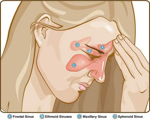
Paranasal sinuses:
Paranasal sinuses are air-filled spaces present within the skull bone around the nasal cavity.
Function:
- Make the skull lighter
- Add resonance to voice
- Air conditioning
Four paranasal sinuses
- Maxillary air sinus
- Frontal air sinus
- Anterior, Middle and posterior ethmoidal air sinus
- Sphenoidal air sinus
Artery supply of nasal septum
- Anterosuperior part
By anterior and posterior ethmoidal arteries
- Anteroinferior part
By the superior labial branch of the facial artery.
- Posterosuperior part
By sphenopalatine artery. It is the main artery.
- Posteroinferior part
By branches of the greater palatine artery.
Kiesselbach's plexus or Little’s area
It is an area in the anteroinferior part of the nasal septum where several vessels supplying the nasal cavity have anastomoses forming a vascular plexus.
This is a common site of nose bleeding.
Bleeding from the nose is called epistasis.
Artery supply of lateral wall of the nasal cavity
1. The anterosuperior part is supplied by the anterior ethmoidal artery & posterior ethmoidal artery
2. The anteroinferior part is supplied by branches from the facial artery
3 The posterosuperior part is supplied by branches of the sphenopalatine artery
4 The posteroinferior part is supplied by branches of the greater palatine artery
(which pierces the perpendicular plate of palatine bone and passes up through the incisive fossa.)
Nerve supply of frontal air sinus
- Supraorbital nerve, the branch of the frontal nerve of the ophthalmic nerve.
Nerve supply of maxillary air sinus
- Posterior superior alveolar nerves from the maxillary and
- Anterior and middle superior alveolar nerves from the infraorbital nerve.
Note:
(Maxillary nerve carries sensation from the maxilla, skin over maxilla, maxillary air sinus, and upper jaw)
Development of paranasal sinuses
The sinuses are in the primitive state, or even absent at birth.
They enlarge rapidly during the ages of 6 to 7 years.
They develop due to the enlargement of pneumatic bone.
Maxillary air sinus is the 1st sinus to develop.
Frontal and ethmoidal sinuses develop after birth.
How is maxillary hiatus reduced in size during living conditions?
How maxillary hiatus is covered by bone?
- Above by
Descending process of lacrimal bone
Uncinate process of ethmoid bone
- From behind by a perpendicular plate of palatine bone
- From below by inferior nasal conchae
Clinical anatomy
|
Paranasal sinuses add resonance to the voice. In common cold and sinusitis, the voice is altered.
|
|
The infection of the sinus is called sinusitis.
Sinusitis may occur due to air pollution.
|
|
Maxillary sinusitis is more common because
It may be infected from the nose or from various tooth caries.
Drainage of the sinus is difficult because its ostium(opening) lies at a higher level than the floor
|
|
Common cold or rhinitis is the most common infection of the nose.
|
|
The sphenopalatine artery is the artery of epistaxis.
Little's area on the nasal septum is a common site of bleeding from the nose or epistasis.
|
|
Hypertrophy of the mucosa over the inferior nasal concha is a common feature of allergic rhinitis, which is characterized by sneezing, nasal blockage, and excessive watery discharge from the nose.
|
|
Pain from the ethmoid & frontal air sinus may be referred to as forehead, as both are supplied by the ophthalmic division of the trigeminal nerve.
|
|
Pain of maxillary sinusitis may be referred to upper teeth and infraorbital skin as all these are supplied by the maxillary nerve.
|
Comments (0)