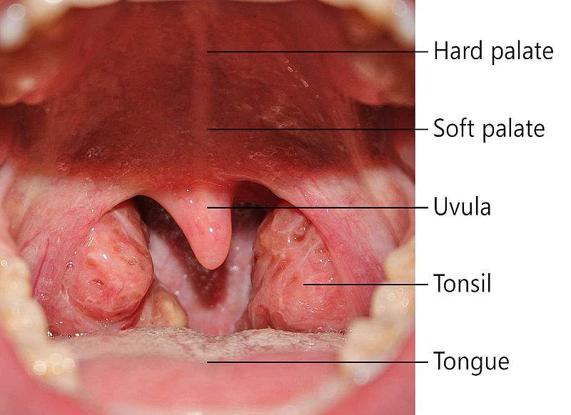
Palatine Tonsil
Palatine Tonsil (commonly called tonsil) is the mass of non-encapsulated lymphoid follicles/nodules present beneath the mucous membrane.
Belong to mucous-associated lymphoid tissue (MALT)
Present on the lateral wall of the oropharynx situated in the tonsillar fossa.
Tonsillar fossa Boundary
- Anteriorly by palatoglossus arch and
- Posteriorly by palatopharyngeal arch
- Superiorly by the soft palate
- Inferiorly by posterior 1/3rd of tongue
Features of Palatine Tonsil
It has two surfaces
- Medial surface
- Lateral surface
The medial surface is covered by non-keratinized stratified squamous epithelium.
Medial surface has 12-15 crypts.
The largest one is called intratonsillar cleft.
The lateral surface is covered by a capsule which is an extension of the pharyngobasilar fascia.
Tonsillar Bed Formation
(From inward to outward)
- The pharyngobasilar fascia
- The superior constrictor & palatopharyngeus muscles
- The buccopharyngeal fascia
In the lowest part,
- The styloglossus & glossopharyngeal nerve
Arterial Supply of Tonsil
Main source: Tonsillar branch of the facial artery.
Additional sources:
- Ascending palatine branch of the facial artery
- Dorsal lingual branches of the lingual artery
- Ascending pharyngeal branch of the external carotid artery
- The greater palatine branch of the maxillary artery
Venous Drainage
Drain to facial vein or palatine vein
Nerve supply of tonsil
- Glossopharyngeal nerve
- Lesser palatine nerve, branch of pterygopalatine ganglion
Development of tonsil
- Epithelium over the tonsil develop from 2nd pharyngeal pouch (endodermal in origin)
- Lymphatics develop from mesoderm
Histology of palatine tonsil
The palatine tonsil is situated at the oropharyngeal isthmus.
Its oral aspect is covered with non-keratinized stratified squamous epithelium, which dips into underlying tissue to form the crypt in the form of nodules.
The structure of tonsil is not differentiated into
- Outer cortex
- Inner medulla
Tips
The function of crypt is to capture bacteria and other debris.
Function:
Bacteria trapped in crypts may induce the formation of antibodies by lymphatic follicles.
It forms the lateral part of Waldeyer’s ring which guards enter to digestive & respiratory passage.
Waldeyer’s Lymphatic Ring:
Aggregated collection of lymphoid tissues is distributed in the pharyngeal mucosa and situated at the junction of the upper respiratory & upper digestive tract in the form of a ring is called Waldeyer’s lymphatic ring.
Formation:
- Posteriorly & above - Nasopharyngeal tonsil (Adenoid)
- Superolaterally presence of Tubal tonsil (Elevated area just above the opening of an auditory tube)
- Inferolaterally - Palatine tonsil (so-called tonsil)
- Inferior on the dorsum surface of posterior 1/3rd of the tongue - lingual tonsil.
Clinical Anatomy
|
1) Quinsy (Peri-tonsillar abscess):
Abscess (pus) formation in the peritonsillar area (between the lateral surface of the tonsil and superior constrictor muscle)
|
|
2) Tonsillectomy is the surgical removal of infected tonsils.
Tonsils are often sites of a septic focus.
|
3) The excessive hypertrophy of the nasopharyngeal tonsil (adenoid) can block the posterior nasal opening making it difficult to breathe (mostly found in children).
These tonsils are large in children. They retrogress after puberty.
Hypertrophy of the tubal tonsil may occlude the auditory tube leading to middle ear problems.
|
4) Tonsillitis may cause referred pain in the ear as the glossopharyngeal nerve supplies both ears.
|
5) Tonsils have only efferent lymph vessels, but no afferent vessels.
|

Comments (0)