Larynx
Pathway for air
Air- Nose- Nasal cavity
|
Posterior nasal apparatus
|
Nasopharynx
Oropharynx
|
Laryngeal inlet
Larynx
|
Below the lower border of the cricoid cartilage
The larynx is continuous as the Trachea
Larynx
Extension: From the inlet of the larynx to the lower border of the trachea
Vertebral level: C3 to C6
At puberty, the male larynx grows rapidly and becomes larger than the female, producing the prominent structure on the anterior of thyroid cartilage known as the Adam’s apple which makes the voice of the male louder and low-pitched.
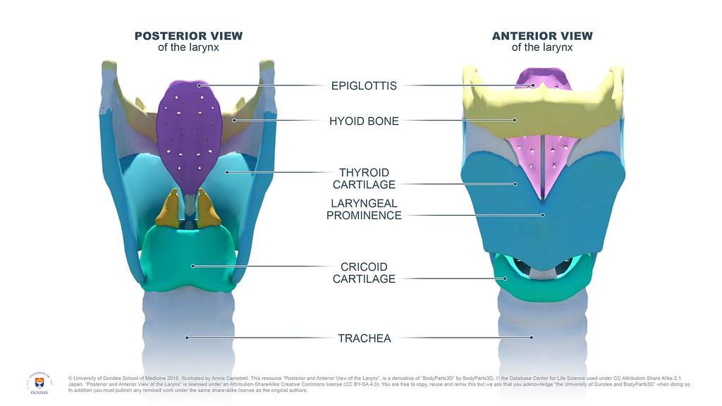
Cartilages of Larynx
Total 9 cartilages among which three are unpaired and three are paired.
Unpaired cartilage
- Thyroid cartilage (V-shaped)
- Cricoid cartilage (ring-like)
- Epiglottis (leaf-like)
Paired cartilages
- Arytenoid
- Corniculate
- Cuneiform
Thyroid cartilage:
Largest V-shaped cartilage
Consist of the Right and Left Lamina which fused anteriorly making a prominent structure called Adam’s Apple in male.
Importance landmark:
The common carotid artery is bifurcated into the external and internal carotid artery, from the upper border of the thyroid cartilage.
Cricoid cartilage:
Ring like shape
The anterior part is narrow(arch) & posterior part (lamina) is broad.
Importance landmark from cricoid cartilage
Cricoid cartilage corresponds to the level of 6th cervical vertebrae
Form the lower border of cricoid cartilage:
- Larynx is continuous below as the Trachea
- Laryngopharynx is continued below as the esophagus
Epiglottis
Leaf-like shaped
Anteriorly attached to the tongue by 3 fold of mucous
- Median glossoepiglottic fold
- Two lateral glossoepiglottic folds
Anterior Surface as related to oral cavity lined by non-keratinized stratified squamous epithelium.
Posterior surface is covered by a smooth mucous membrane lined by respiratory epithelium (pseudostratified ciliated columnar epithelium)
Epiglottis is attached to arytenoid cartilage by aryepiglottic fold.
Aryepiglottic muscle helps to close the inlet of the larynx during swallowing.
Epiglottis forms the anterior boundary of the laryngeal inlet.
Function of epiglottis:
During deglutition, it moves forward and upward by longitudinal muscles of the pharynx & is squeezed between the base of the tongue & larynx.
So food bolus slips over its anterior surface as it bends back over the laryngeal inlet.
Arytenoid cartilage
Pyramidal shaped
Rest on the lamina of the cricoid cartilage on its upper border.
Attachment
- Vocal process:
Vocal fold and vocalis muscle is attached
- Above the vocal process:
Vestibular fold is attached
- Muscular process Attachment:
-Posterior cricoarytenoid muscle
-Lateral cricoarytenoid muscle
-Transverse arytenoid muscle
Corniculate cartilage:
Lie on the apex of arytenoid cartilage just the posterior part of the aryepiglottic fold
Cuneiform cartilage:
Lie in the aryepiglottic fold ventral to the corniculate cartilage
Histology of laryngeal cartilage
Hyaline cartilage
- Thyroid cartilage
- Cricoid cartilage
- Basal part of arytenoid cartilage
They ossify after the age of 25 years
Elastic cartilage
- Epiglottis
- Processes of arytenoid
- Corniculate
- Cuneiform
They don’t ossify throughout the life
Laryngeal joint
Cricothyoid joint (Synovial joint)
- Between the inferior cornea of the thyroid cartilage and the side of the cricoid cartilage.
- They permit tension and relaxation of vocal cord
Cricoarytenoid joint (Synovial joint)
- Between the base of the arytenoid cartilage and the upper border of the lamina of the cricoid cartilage.
- They permit the adduction and abduction of vocal cords.
[Laryngeal ligaments and Membranes]
Extrinsic ligaments & membrane
1) Thyroid membrane:
Connects the thyroid cartilage to the hyoid bone.
The membrane is pierced by
- Internal laryngeal nerve, branch of superior laryngeal nerve of the vagus nerve and
- Superior laryngeal artery, branch of superior thyroid artery of external carotid artery
2) Hyoepiglottic ligament connects the epiglottis to hyoid bone
3) Cricotracheal ligament connects cricoid cartilage to the upper end of the trachea.
Intrinsic membrane and ligament of larynx
1) Quadrate membrane:
- Extends from epiglottis to arytenoid cartilage
- Upper free border thicken to form aryepiglottic fold
- Lower free border thickened to form vestibular ligament which after surrounded by mucus membrane form vestibular fold
2) Vestibular ligament
3) Conus Elasticus
- Extends from anterior arch of the cricoid cartilage to the inner aspects of thyroid cartilage (anteriorly) and arytenoid cartilage (posteriorly)
- Upper free margin of conus elasticus thicken to forms vocal ligament which after surrounded by mucus membrane form vocal fold.
4) Vocal ligament
| Within the cavity of the larynx, there are two folds of mucus membrane on each side. |
Vestibular fold (False vocal cord):
Fold of mucus membrane of larynx around the vestibular ligament (which is formed by the lower free margin of quadrate membrane) is called vestibular fold. |
|
Vocal Fold (True vocal cord)
The fold of mucus membrane of larynx around the vocal ligament (which is formed by upper free margin of conus elasticus) is called vocal fold.
- Vocal fold is attached anteriorly to the thyroid cartilage and posterior to the vocal process of the arytenoid cartilage.
- Lateral to the vocal ligament lies vocalis muscle.
|
Within The Cavity of Larynx
The cavity of larynx extends from the inlet of the larynx to the lower border of the cricoid cartilage.
Laryngeal inlet bounded on each side by piriform fossa.
Boundary of the inlet of the larynx:
- Anteriorly by epiglottis
- On each side by aryepiglottic fold [LATERAL TO THIS - PIRIFORM FOSSA]
- Posteriorly by inter arytenoid fold
Piriform fossa
Piriform fossa is the fossa which is present in the lateral wall of the Laryngopharynx.
Or can be said as fossa present on either side of the laryngeal inlet.
Boundary of piriform fossa:
- Medially- aryepiglottic fold
- Laterally - medial surface of thyroid cartilage & thyrohyoid membrane.
Importance:
- Beneath the mucosa of the fossa there lie internal laryngeal nerve.
Removal of foreign body from this fossa may damage the nerve.
- Use by smugglers to hide precious materials (so-called as Smuggler fossa)
Within the cavity of the larynx, there are two folds of mucus membrane on each side.
- The upper fold is vestibular fold (false vocal cord) &
Lower fold is the vocal fold (True vocal cord)
- Space between the right and left vestibular folds is Rima Vestibuli.
- Space between the right and left vocal folds is Rima glottidis.
[Rima is the narrowest part of the larynx.]
Subdivision of laryngeal cavity
The vestibular and vocal fold divides the cavity of the larynx into three parts
- Vestibule of the larynx [Supraglottis]
The part above the vestibular fold and below the aryepiglottic fold
- Ventricle of larynx [Sinus of larynx]
The part between the vestibular and vocal folds
- Infraglottis
The part below the vocal folds
[ The extension of the sinus of the larynx is called the saccule of the larynx which contains mucous glands which helps to lubricate the vocal folds.
It is often called oil can of larynx.]
Lining epithelium of the mucous membrane of the larynx
- Anterior surface of epiglottis &
upper half part of the posterior surface of the epiglottis
- Upper part of the aryepiglottic folds
- Vocal folds
Lined by non-keratinized stratified squamous epithelium
- Rest of other mucous membranes of the larynx
Lined by respiratory epithelium (pseudostratified ciliated columnar epithelium)
Tips
As the anterior part of the epiglottis and the aryepiglottic fold is related to food passage, they need to be highly protective. So that pressure of food could tear out the mucous membranes.
That’s why lined by stratified (multiple layers) epithelium.
Similarly, Stratified (multi-layer cells) is needed to provide extra protection to the vocal cord against the foreign bodies which may enter the larynx accidentally.
[Intrinsic muscles of Larynx]
Cricothyroid: the only muscle which lie outside
-Posterior cricoarytenoid
-Lateral cricoarytenoid
-Transverse arytenoid
Oblique arytenoid
-Aryepiglottic
-Thyroepiglottic
Thyroarytenoid
-Vocalis
Muscles acting on the larynx
| Movement |
Muscle |
| Elevation of larynx |
Thyrohyoid, mylohyoid |
| Depression of larynx |
Sternothyroid, sternohyoid |
| Opening inlet of larynx |
Thyroepiglottic |
| Closing inlet of larynx |
Aryepiglottic |
| Abductor of vocal cords |
Posterior cricoarytenoid only |
| Adductor of vocal cords |
Lateral cricoarytenoid,
transverse and oblique arytenoids
|
| Tensor of vocal cords |
Cricothyroid |
| Relaxor of vocal cords |
Thyroarytenoid and vocalis |
Tips
The vocal process and muscular processes move in opposite directions.
Any muscle which pulls the muscular process medially, pushes the vocal process laterally, resulting in the abduction of vocal cords.
This is done by only one pair of muscle, the posterior cricoarytenoid.
Muscles that pull the muscular process forward, also laterally will push the vocal process medially causing the adduction of vocal cords.
This is performed by lateral cricoarytenoid and transverse arytenoid.
Adduction & Abduction - Cricoarytenoid joint
Tension & relaxation of vocal cord - Cricothyroid joint
Blood supply of the larynx
Arterial supply
Up to vocal folds:
By the superior laryngeal artery, branch of superior thyroid artery of external carotid artery
Below the vocal fold:
By the inferior laryngeal artery, branch of inferior thyroid artery
Tips:
Inferior thyroid artery, branch of thyrocervical artery which is the branch of 1st part of subclavian artery
Mnemonic for branch of subclavian artery: VIT C D
Venous Drainage:
Up to the vocal fold:
Superior laryngeal vein drains into superior thyroid vein which drains to the internal jugular vein
Below the vocal fold:
By inferior laryngeal vein drain into inferior thyroid vein which then drain into the left brachiocephalic vein.
Nerve supply of larynx
Motor supply:
All the intrinsic muscles of the larynx are supplied by the recurrent laryngeal nerve
Except the cricothyroid - supplied by the external laryngeal nerve.
Sensory supply:
- Mucous membrane above the vocal folds
Supplied by internal laryngeal nerve
- Mucous membrane below the vocal folds
Supplied by the recurrent laryngeal nerve
Tips
Branches of vagus nerve
1) Superior laryngeal nerve
- Internal laryngeal nerve (sensory nerve)
_Carry taste sensation from the posteriormost part of the tongue and epiglottis region.
_Sensory supply above the level of the vocal cord
- External laryngeal nerve (motor nerve)
Supply only cricothyroid muscle
2) Recurrent laryngeal nerve (mixed nerve)
- Motor supply to all the intrinsic muscles of the larynx except cricothyroid
- Sensory supply to larynx below the level of vocal fold
Course of Right & Left Recurrent laryngeal nerve varies
- The right recurrent laryngeal nerve hooks around the right subclavian artery and then ascend upward.
- Left recurrent laryngeal nerve hooks around the arch of the aorta and then ascend upward.
Development of larynx:
The larynx is developed from the cephalic part of Laryngo-tracheal tube which has been developed from the respiratory diverticulum which has grown the ventral wall of the pharyngeal part of the foregut
(Endodermal in origin)
Cartilages of larynx develop from 4th & 6th branchial arches.
(mesodermal in origin)
Clinical Anatomy
|
When any foreign object enters the larynx, a cough reflex occurs to expel the object.
However, damage to the internal laryngeal nerve produces anesthesia of the mucous membrane in the supraglottic part of the larynx, so that foreign bodies can readily enter it.
|
|
Fishbone may get stuck in the vallecula or piriform fossa.
Often theses bones just scratch the mucosa on their way down, and the person gets feeling of foreign body sensation, due to a dull visceral pain caused by the scratch, which is carried out by internal laryngeal nerve (sensory Nerve)
|
|
On should be careful while taking out the stuck fish bones, because below the mucosa of the piriform fossa, there is an internal laryngeal nerve.
If in any way, internal laryngeal nerve-damaged, then it produce anesthesia of the mucous membrane and cough reflex is not generated.
So foreign bodies can easily enter into the larynx.
|
|
The infection of the larynx is called laryngitis, which causes hoarseness of voice (roughness of voice).
|
|
Misuse of the vocal cords may produce nodules on the vocal cords called Singer’s nodules or Teacher’s nodule.
|
|
The mucus membrane of the larynx is supplied by the X nerve through superior laryngeal or recurrent laryngeal nerves.
So laryngeal tumors may also cause referred pain in the ear partly supplied by auricular branch of X nerve.
|
|
Piriform fossa lies between the quadrate membrane and the medial surface of the thyroid cartilage.
It is traversed by the internal laryngeal nerve.
|
|
Piriform fossa is used to smuggle out precious stones, diamonds, etc.
Thus, called as smuggler’s fossa.
|
Posterior cricoarytenoid is the only abductor of the vocal cord and so it is a life-saving muscle.
|
Comments (0)