Embryology Notes Up to Gametogenesis
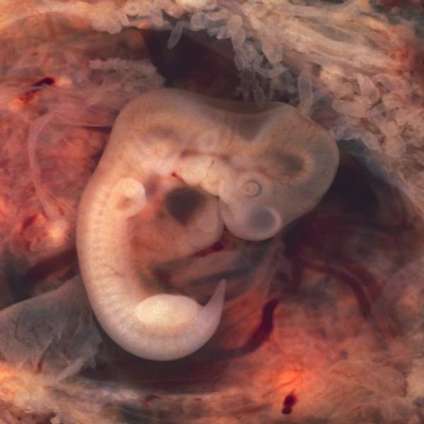
Embryology: It deals with the prenatal stages of development from the fertilization of the ovum to the birth of a new individual.
The zygote by a series of changes – cell division, growth, differentiation, cell migration, delamination, and folding.
Ontogeny: Complete the life cycle of an organism. It consists of prenatal development and postnatal growth and maturity then aging, sanity, and death.
Phylogeny: Encompasses the evolution or ancestral history of a group of organisms.
Ascending order of vertebrates -fish - amphibians - reptiles - birds - mammals.
The basic process of development: Two processes
- Growth
- Differentiation
Growth means an increase in bulk. Which may take place in one of the three ways
- Multiplicative: cell division causes an increase in cell number. It occurs by mitotic cell division, obstructed in most of the tissue and organs in prenatal life.
- Auxetic: by increasing cell size. E.g oocyte.
- Accretionary: by increasing accumulation of intracellular substance .e.g bones, cartilages (connective tissue).
Differentiation: It is a complicated process in which a group of cells assume special characteristics and are assigned specific functions.
Stages:
- Totipotent: After the formation of the zygote each of the cells derived from the first cleavage division possesses a totipotent character. And each cell produces a separate embryo. Totipotent nature may be retained up to the 8 cells of cleavage division.
- Plastic phase
- Chemo differentiation
- Histo differentiation
- Organogenesis
- Functional differentiation
Cell cycle: The period of time between the birth of a cell as a result of the division of its parent cell and its own division to produce two daughter cells is known as the cell cycle. Or The alteration between mitosis and interphase is called the cell cycle.
Events of cell cycle
- Karyokinesis-nuclear division
- Cytokinesis-cytoplasmic division
Where does mitosis occur?
Mitosis occurs in all somatic cells and immature germ cells e.g. spermatogonia, and oogonia.
Where does meiosis occur?
Meiosis occurs in the primary spermatocyte, primary oocyte, sec. Spermatocyte, sec. Oocyte.
Prophase :
- Chromosome begins to shorten, thicken, and recognizable.
- Each chromosome split longitudinally into two chromatids except at the centromere.
- Outside the nucleus, centriole begins to separate moving to opposite poles of the cell.
- Central spindle formed by continuous elongation of microtubules between the centrioles and astral rays form.
- The nuclear membrane and nucleolus disappear.
Metaphase:
- Chromosomes move to the equator of the spindle.
- Chromosomes are attached by their centromeres to spindle microtubules.
Anaphase:
- Centromere split longitudinally.
- Two chromatids separate, to form two new chromosomes and move toward each pole
Telophase:
- The daughter chromosomes are collected at each end of their spindle and are involved by the nuclear membrane and the nucleolus reappear.
Before mitosis, the chromosome number is 46. After meiosis, the chromosome number is 23.
Non-disjunction :(VVI)
After the splitting of chromosomes, one or more chromosomes fail to migrate properly. Due to the abnormal function of the spindle apparatus. This phenomenon is known as non-disjunction.
As a result of trisomy, monosomy occurs.
Meiosis l
A. Prophase l
- Laptotene
- Chromosomes begin to shorten, thicken, and recognizable and attached to the nuclear membrane.
- Chromosomes appear as beads.
- Zygotene
- The pairing of chromosomes i.e. synapsis occur
- Chromosomes have come together side by side in homologous pairs.
- Pachytene
- Pairing is complete and tetrad formation occurs.
- Crossing over also occurs.
- Diplotene
- Chiasmata form where crossing over has occurred.
- Chiasmata form where crossing over has occurred.
- Diakinesis
- Nucleoli and nuclear membranes disappear.
B. Metaphase l
Chromosome pairs are attached to the spindle and are arranged in an equatorial plane.
C. Anaphase l
Paired chromosomes separate and move towards the pole.
D. Telophase l: Like mitosis
The purpose of the result of meiosis:
- Genetic variability is enhanced through crossing over.
- The haploid number of chromosomes forms. So that at fertilization the diploid number is restored.
Go phase: In this phase, further cell division does not occur and cells do not enter the S phase
Permanent cells: Nerve cells and skeletal muscle cells.
Before meiosis: DNA-4n
After meiosis l : DNA-2n
After meiosis ll: DNA-1n
Gametogenesis
Spermatogenesis: The process of formation of sperm is called spermatogenesis.
Events:
- Spermetoctosis
- Meiosis
- Spermiogenesis
Spermatocytosis: It is the process by which spermatogonia are transformed into primary spermatocytes.
Meiosis: It is the process by which primary spermatocytes transform into spermatids.
Spermiogenesis: The series of changes resulting in the transformation of spermatids into spermatozoa is called spermiogenesis.
The changes that occur in spermiogenesis include:
- Condensation of the nucleus.
- Formation of the acrosome.
- Formation of the neck, middle piece, and tail.
- Shedding off extra cytoplasm. (That is called residual body)
Oogenesis: It is the process by which the ovum is formed from oogonia.
Process of oogenesis:
Primordial germ cell (develop oogonia and spermatogonia) arises from the endoderm of the yolk sac and reach the gonads (testes, ovary), where it differentiates into oogonia. Oogonia undergo mitotic divisions and by the end of the third month clusters of oogonia are surrounded by a layer of flat epithelial cells known as follicular cells, originating from surface epithelium. Then oogonia enters into mitotic cell division but is arrested in prophase of meiosis l and forms the primary oocyte. Most of the oogonia and primary oocytes become degenerate. All surviving primary oocytes are surrounded by layers of flat epithelial cells. A primary oocyte with its surrounding flat epithelial cells is known as the primordial follicle. Primary oocyte remains in prophase In the diplotene stage and does not finish their meiotic division before puberty is reached.
The total no. Of primary oocyte at puberty is estimated as 4,00,000 and fewer than 500 will be ovulated. At puberty 15-20 follicle begins to mature.
Thereafter primary follicle develops where the primary oocyte surrounded by follicular cells changes to form flat to cuboidal epithelial. And proliferate to form stratified epithelium of granulosa cells. Surrounding stroma (connective tissue) cells proliferate to form theca follicle. Finally, a mature Graafian follicle develops.
Graffian follicle:
A mature Graafian follicle consists from outside to inside :
- Theca externa
- Theca Interna
- Stratum granulosum
- Zona pellucida
- Perivitelline space with first polar body
- Oolema
- Ooplasm
- Germinal vesicle

Comments (0)