Lung
Introduction
- It is the main organs of respiration.
- It is situated in the thoracic cavity on either side of the mediastinum.
- It is enclosed in the pleural sac.
- It is pyramidal in shape.
- It helps in the exchange of gaseous between inspired air and blood.
- The weight of the right lung is 700gm whereas the weight of the left lung is 650g.
- The right lung has three lungs whereas the left lung has two lobes.
- The lungs are attached to the trachea by pulmonary bronchi.
- The lungs are attached to the heart by the pulmonary vessels.
- The newly born baby who lives in a clean environment has rosy-pink color lungs.
- Similarly, those who live in a polluted environment have brown or black color lung.
- The lungs of an adult are spongy in texture and crepitate in touch.
- But the lungs of the fetus are solid and not crepitate in touch because of the absence of air in alveoli.
External features
- Apex
- Base
- Three borders ( anterior, posterior, and inferior)
- Two surfaces ( costal and medial)
Apex
- It is a rounded superior end of the lung.
- It is covered by cervical pleura and supra pleural membrane.
Relation
Anteriorly:
- Subclavian artery
- Internal thoracic artery
- scalenus anterior
posterior:
- The neck of the first rib.
- The ventral ramus of the first thoracic nerve.
- First posterior intercostal artery
- First posterior intercostal vein
- Sympathetic chain
Base
- it is a lower semilunar concave surface that rests on the dome of the diaphragm.
- It is also called a diaphragmatic surface.
Relation
- On the right side: it is separated from the liver by the right dome of the diaphragm.
- On the left side: it is separated from the spleen and fundus of the stomach by the left dome of the diaphragm
Border
1. Anterior border
Right lung
- The anterior border of the right lung is vertical.
Left lung
- A wide cardiac notch is present in the anterior border.
- Cardiac notch is occupied by the heart and pericardium.
- In the area of cardiac notch heart and pericardium is not covered by lung.
- That’s why this area is responsible for cardiac dullness.
- Tongue shaped projection present below the cardiac notch is called lingula.
Posterior Border
- It is thick and rounded.
- It extends from the spine of the C7 vertebrae to the spine of the T10 vertebra.
Inferior border
- It is semilunar in shape in shape and separates the base from the costal and medial surface.
Surface
Two types
1. Costal surface
2. Medial surface
- Small posterior Vertebral part
- Large anterior Mediastinal part
Costal surface
- It is large, smooth, and convex.
- costal pleura and endo-thoracic fascia cover this surface.
The number of ribs related to this surface
- Upper 6 ribs related in the midclavicular line.
- Upper 8 ribs in the midaxillary line.
- Upper 10 ribs in the scapular line.
Medial surface
Vertebral part
Relation
- Vertebral column
- Posterior intercostal vessels
- Greater and lesser splanchnic nerve
Mediastinal part
- Hilum present in this part.
- It is related to mediastinal structures such as heart, great vessel, and nerves.
Hilum
- it is the area of lungs where various structures enter and exit.
The structure which enters
- Two principle bronchus ( where gases are exchanged). Right principle bronchus is divided into eparterial and hypaterial bronchus. Whereas left principle bronchus does not divide.
- Pulmonary artery
- Bronchial artery
- Pulmonary plexus
The structure which exits.
- Pulmonary vein
- Bronchial vein
- Lymphatic vein
Structures forming the right surface of the mediastinal
- The right mediastinal surface mainly consists of the right atrium.
- Above the right atrium, superior vena cava and right brachiocephalic vein are present. Inferior vena cava also present.
- Trachea and esophagus are behind the mediastinum.
- The azygos vein(a large venous channel), runs upward along the side of the vertebral column and make an arch over the root of the right lung to end into superior vena cava.
- Right, the phrenic nerve run to the diaphragm passing to the superior vena cava from above to downward in front of the root of the lung.
- Vagus nerve lies against the right side of the trachea and travels behind the root of the lung to break and form posterior pulmonary plexus and oesophageal plexus.
- Sympathetic trunk runs in the paraventricular gutter. The splenic nerve leaves its lower half to pierce the crura of the diaphragm to reach the abdomen medially.
The left surface of the mediastinum
- The left ventricle and aorta are the main structure of the surface.
- Aorta ascends at first, arches over the left lung root, and then descends behind the lung root.
- Three great vessels present ( brachiocephalic trunk, left common carotid artery, and left subclavian vein) arise from the aortic arch and ascend up to reach the root of the neck.
- Esophagus shifts to the right, behind the heart and gently cross the line of descending aorta.
- Left phrenic nerve crosses the aortic left side, passing root of the lung and runs down superficial to left ventricle to reach the diaphragm.
- The left vagus nerve is held away from the trachea by the aortic arch. It gives a laryngeal branch which hooks under the aortic arch, ascending into the tracheoesophageal groove. Below aortic arch, vagus nerve runs behind the lung root and breaks up into posterior pulmonary and oesophageal branches.
- Sympathetic trunk and splanchnic nerve positions are similar to those right side.
Root of lung
- It is a short broad pedicle connecting the medial surface of the lung with mediastinum.
- It consists of structures entering and leaving the lung at the hilum.
- It is surrounded by tubular sheath derived from the mediastinal pleura.
Components of the root of the lung
Name of some structure consisting root of the lung.
- Principal bronchus in the left lung, eparterial and hyparterial bronchi in the right lung.
- Pulmonary artery
- Pulmonary veins ( Two in Number)
- Bronchial arteries( one on right side two on the left side)
- Bronchial veins
- Lymphatics of the lung.
- Anterior and posterior pulmonary plexus of the nerves.
Hilum of lung
- It is the area on the mediastinal surface of the lung through which structures enter or leave the lung.
Note:
- The root of the lung lies opposite to the bodies of the T5, T6, and T7 vertebrae.
- Arrangement of structure in the root of the lung
From before backward
- Pulmonary vein
- Pulmonary artery
- Bronchus
From the above downward, it differs on two sides.
Right side
- Eparterial bronchus
- Pulmonary artery
- Hyparterial bronchus
- Inferior pulmonary vein.
Left side
- Pulmonary artery
- Left principle bronchus
- Inferior pulmonary vein
Relation of the root of the lung
Anterior
- Phrenic nerve
- Anterior pulmonary plexus
- Superior vena cava ( on the right side only)
Posterior
- Vagus nerve
- Posterior pulmonary plexus
- Descending thoracic aorta ( on the left side only)
Superior
- Arch of the azygos vein (on the right side only)
- Arch of the aorta (on the left side only)
Inferior
Lobes and fissures
Right lung
Lobes= Three
- Superior
- Middle
- Inferior
Fissures= Two
- An Oblique fissure
- A Horizontal fissure
Left lung
Lobes = 2
- Superior
- Inferior
Fissure = 1
- Oblique fissure
Oblique fissure
- It runs obliquely downward and forward crossing the posterior border.
- It separates the superior and middle lobes from the inferior lobe in the right lung.
- It separates the superior lobe from the inferior lobe in the left lung.
Horizontal fissure
- It is only present in the right lung.
- It starts from the oblique fissure at the midaxillary line and runs horizontally forward to the anterior border of the lung.
- It separates the superior and middle lobes.
- The internal structure of the lung
Intrapulmonary bronchial tree
- The lung is mainly made up of the intrapulmonary bronchial tree.
- It is concerned with the condition of air to and fro from the lung.
Pulmonary unit
- It is concerned with the gaseous exchange within the lung.
It consists of
- Alveolar ducts
- Atria
- Air saccules
- Alveoli
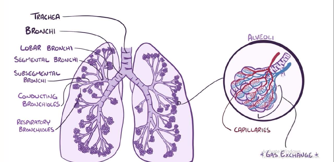
Bronchial tree
The bronchial tree consists of
- The principal bronchus
- Lobar bronchi
- Terminal bronchioles
- Respiratory bronchioles
Principal bronchi
- At the level of the T4 vertebra, the trachea divides outside the lungs into two primary bronchi
- Right and left primary bronchi for both lungs
Write difference from the book
Lobar bronchi
- When the principal bronchus enters inside the right lung it divides into three lobar bronchi, one for each lobe.
- Similarly, when the principal bronchus enters inside the left lung it divides into two lobar bronchi, one for each lobe.
Tertiary bronchi/Segmental Bronchi
- Lobar bronchi divide into segmental bronchi, one for each bronchopulmonary segment.
- Segmental bronchi divide into very small bronchi called terminal bronchioles.
- The terminal bronchioles give off respiratory bronchioles.
- Respiratory bronchioles aerate a small portion of the lung called pulmonary units.
Notes
- The respiratory bronchiole represents the transitional part between the conducting and respiratory portions of the respiratory system.
Alveoli
- These are specialized sac-like structures that form a greater part of the lungs.
- They are the main sites for the gaseous exchange of oxygen and carbon dioxide between the inspired air and blood.
Bronchopulmonary segments
- These are well defined, wedge-shaped sectors of the lung, which is aerated by tertiary bronchi.
Characteristics features
- It is the subdivision of the lung.
- It is pyramidal in shape with the apex directed toward the hilum and base toward the surface of the lung.
- It is surrounded by connective tissue.
- It is aerated by the segmental bronchus.
- Each segment has its own artery
- Each segment has its own lymphatic drainage and autonomic nerve supply.
- Drained by intersegmental veins.
- Surgically resectable.
Note:
- It is the smallest well defined.
- It is anatomically independent.
- It is also known as functional and surgical units of the lungs.
- Because of all those reasons they can be isolated and removed without affecting adjacent regions.
Table of bronchopulmonary segments from the book
Artery supply of the lungs
- Bronchial arteries
- Pulmonary artery
Bronchial artery
- It supplies nutrition to the bronchial tree and pulmonary tissue.
Right lung= it is supplied by one bronchial artery which arises from the third posterior intercostal artery
Left lung= it is supplied by two bronchial arteries which arise from descending thoracic aorta
Pulmonary artery
- It supplies deoxygenated blood to the lungs.
- There is one pulmonary artery for each lung.
- These are the branches of the pulmonary trunk.
Venous drainage
- Bronchial veins
- Pulmonary veins
Bronchial vein
- It drains the deoxygenated blood from bronchial tree and pulmonary tissue.
- Right bronchial veins drain into azygos veins.
- The left bronchial veins drain into the hemiazygos veins or the left superior intercostal veins.
Pulmonary veins
- The pulmonary veins drain the oxygenated blood from the lungs.
- There is two pulmonary veins into two sides.
Nerve supply
- It is supplied by both parasympathetic fiber and sympathetic fiber.
- Both provide motor supply to the bronchial muscles and secretomotor supply to the mucus glands of the bronchial tree.
Parasympathetic fibers
- Derived from the vagus nerve
- It causes bronchoconstriction, vasodilation and increased mucus secretion.
Sympathetic fibers
- derived from the T2 to T5 spinal segment.
- It causes bronchodilatation, vasoconstriction, decreased mucus secretion.
Comments (0)