Gram Staining Procedure
3 years ago 28464
Gram staining is a laboratory procedure used to differentiate bacterial species based on the properties of their cell walls.
Here are the step-by-step instructions for the Gram staining procedure:
Materials required for the gram staining procedure are:
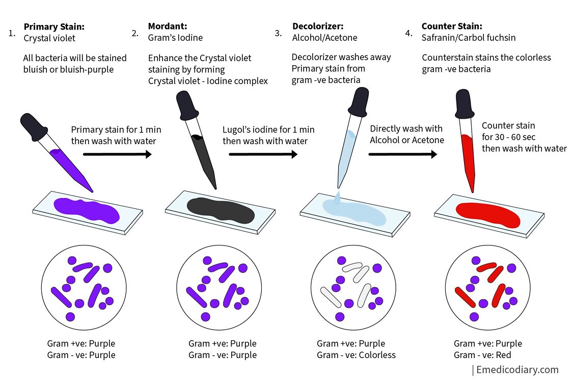
There are 4 basic steps in Gram Staining Procedure
Primary staining is done with Crystal violet for 1 minute, then washed with water.
Lugol's Iodine is added as Mordant for 1 minute, then washed with water.
A decolorizing agent (Alcohol or Acetone) is added.
Counterstaining is done by Carbol fuchsin or Safranin for 30-60 seconds, then washing with water.

Interpretation of Gram staining procedure
Gram staining is a differential staining technique used to distinguish between different types of bacterial cells based on the properties of their cell walls. The principle behind Gram staining is based on the fact that bacteria can be classified into two groups based on their cell wall structure and composition:
1. Gram-positive bacteria: These bacteria have a thick layer of peptidoglycan in their cell walls that retains the Primary stain (Crystal violet stain) without being washout by Decoloizer, thus, they do not take the Counter stain (Carbol fuchsin or Safranin) during the staining process. This gives them a purple color under the microscope.
2. Gram-negative bacteria: These bacteria have a thinner layer of peptidoglycan in their cell walls, as well as an outer membrane that contains lipopolysaccharides. During the staining process, the Primary stain (Crystal violet stain) is washed out of their cell walls by the decolorizing agent, and they take up the Counterstain (Carbol Fuchsin or Safranin). This gives them a pink or red color under the microscope.
The Gram staining procedure involves a series of steps, including staining the bacterial cells with Primary stain (Crystal violet), treating them with a Mordant (Lugol's iodine), Decolorizing with alcohol, and Counterstaining with safranin or Carbol fuchsin. These steps help to differentiate between gram-positive and gram-negative bacteria based on the properties of their cell walls. The Gram stain is widely used in microbiology to help identify different types of bacteria and to guide appropriate treatment decisions.
1. Prepare a bacterial smear: Using a sterile metallic loop, transfer a small amount of bacterial culture onto a clean microscope slide. Spread the culture into a thin, even film on the slide.
2. Air dry the smear: Allow the bacterial smear to air dry completely at room temperature. Do not heat-fix the smear.
3. Heat-fix the smear: Pass the slide through the flame of a Bunsen burner in To and From motion 2-3 times to heat-fix the bacterial cells to the slide.
4. Primary Staining of the smear: Flood the heat-fixed bacterial smear with a Crystal violet stain for 1 minute. Rinse the slide gently with distilled water to remove excess stain.
5. Apply Lugol's iodine: Cover the smear with Gram's iodine solution for 1 minute. This acts as a mordant and helps to fix the crystal violet stain in the bacterial cells.
6. Decolorize the smear: Quickly rinse the slide with 95% ethanol or 70% isopropyl alcohol until no more purple color runs off. This removes the crystal violet stain from some bacteria, making them colorless or "gram-negative". Be careful not to over-decolorize the smear, as this can result in false-negative results.
7. Counterstain the smear: Flood the slide with safranin stain or carbol fuchsin stain for 1 minute. This stains the decolorized bacteria pink or "gram-negative", allowing them to be seen under the microscope.
8. Rinse and dry the smear: Rinse the slide gently with distilled water to remove excess stain, and blot the slide dry with a paper towel.
After completing the above gram staining procedure, observe the stained smear under a microscope using a 100x oil immersion lens. Gram-positive bacteria will appear purple or blue-violet, while gram-negative bacteria will appear pink or red.
Note: Proper fixation and staining techniques are essential for accurate results. It is important to note that not all bacteria will stain well using the Gram stain method. Some bacteria, such as mycobacteria, have unique cell wall structures and require different staining methods i.e. Ziehl Neelsen Stain
Comments (0)