The Diaphragm
The Diaphragm
- The diaphragm of inferior thoracic aperture
- The thoracic outlet is closed by a large dome-shaped flat muscle called the diaphragm.
- It separates the thoracic cavity from the abdominal cavity.
- It is also termed as the thoracic abdominal diaphragm.
- Functionally, the diaphragm is the principal muscle of respiration.
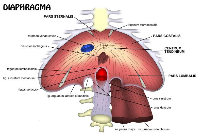
- It is dome-shaped.
It is consists of
- Peripheral muscular part
- The central fibrous part called a central tendon.
Origin
The origin of the diaphragm is divided into three parts,
- Sternal
- Costal
- Vertebral
Sternal part;
- It consists of two fleshy slips.
- Fleshy slips arise from the posterior surface of the xiphoid process.
Costal part;
- On each side, it consists of six fleshy slips.
- Which arise from the inner surface of lower six ribs near their costal cartilages.
Vertebral part;
This part arises by means of
- Right and left crura of the diaphragm
- Five arcuate ligaments.
Crura
Right crus
- It is a vertical fleshy bundle
- It arises from the right side of anterior aspects of L1, L2, L3 vertebrae and inverting intervertebral discs.
Left crus
- It is a vertical fleshy bundle.
- It arises from the left side anterior aspect of L1, L2 vertebrae and the intervening intervertebral discs.
Arcuate ligament
1. Median arcuate ligament
- It is an arched fibrous band stretching between the upper ends of two crura.
2. Medial arcuate ligament
- It is thickened upper margin of the psoas sheath.
- It extends from the side of the body of L2 vertebra to the tip of the transverse process of L1 vertebra.
3. Lateral arcuate ligament
- It is thickened upper margin of fascia covering the anterior surface of quadratus lumborum.
- It extends from the tip of the transverse process of L1 vertebra to the 12th rib.
Note
- The right crus is attached to more number of vertebrae because the right side diaphragm has to contract on the massive liver.
Insertion
- The muscles fibres converge towards the central tendon and insert into its margins.
Features of the central tendon
- It is trifoliate in shape.
- It is inseparably fused with the fibrous pericardium.
- It is located nearer to the sternum than to the vertebral column.
Surfaces
- Superior surface
- Inferior surface
Superior surface
- It is projected on either side as dome-shaped into the thoracic cavity.
- The central tendon is the depressed area between the two domes.
- The superior surface is covered by endothoracic fascia.
Relation of the superior surface
- It is related to bases of right and left pleura on either side.
- The fibrous pericardium is located in the middle.
Inferior surfaces
- It is lined by diaphragmatic fascia and parietal peritoneum.
Relation of the inferior surface
On the right side
- The right lobe of the liver
- Right kidney
- Right suprarenal gland
On left side
- Left lobe of the liver
- Fundus of stomach
- Spleen
- Left kidney
- Left suprarenal gland
Opening of diaphragm
- Major opening
- Minor opening
Major opening
- Vena cava opening
- Oesophageal opening
- Aortic opening
Note
- Contraction of the diaphragm enlarges the caval opening to enhance venous return.
- Contraction of the diaphragm has a sphincteric effect on the oesophageal opening.
- Contraction of the diaphragm has no effect on the aortic opening because strictly speaking it is outside the diaphragm.
Location, shape, and vertebral level of three major openings of the diaphragm
| Opening |
Location |
Shape |
Vertebral level |
| vena caval opening |
In the central tendon slightly to the right of median plane between the central and right posterior leaflet. |
Quardrangular |
T8 |
| oesophageal opening |
Slightly to the left of median plane. |
Oval |
T10 |
| Aortic opening |
In the midline behind the median arcuate ligament. |
Circular |
T12 |
structure passing between three major opening of Diaphragm
| Opening |
Structures passing through |
| Vena caval opening |
- inferior vena cava
- Right phernic nerve
|
| Oesophageal opening |
- oesophagus
- Right and left vagal trunk
- oesophageal branch of left gastric artery
|
| Aortic opening |
from right to left
- Azygos vein
- Thoracic duct
- Aorta
|
Minor opening
- Superior epigastric vessels
- Musculophrenic artery
- Lower five intercostal nerve and vessels
- Subcostal nerve and vessels
- Sympathetic chain
- Greater, lesser and least splanchnic nerves
- Hemiazygos vein
Nerve supply
The diaphragm supplied by
- Right and left phrenic nerve
- Lower five intercostal and subcostal nerve
Phrenic nerve
- The right phrenic nerve provides motor invertion to the right half of the diaphragm up to the right margin of the oesophageal opening
- Left phrenic nerve provides motor invertion to the left of the diaphragm up to the left margin of the oesophageal opening.
- The phrenic nerve provides sensory invertion to the central tendon of the diaphragm, pleura and peritoneum related to it.
Intercostal nerve
- It supplies the peripheral part of the diaphragm.
Artery supply
- Superior phrenic artery
- Inferior phrenic artery
- Pericardiophrenic arteries
- Musculophrenic arteries
- Superior epigastric arteries
- Lower five intercostal and subcostal arteries
Development
The diaphragm develops in the region of the neck from the following four embryonic structures
- Septum transversum
- Pleuroperitoneal membranes
- The dorsal mesentery of oesophagus
- Body wall
Adult derivative
- Central tendon of diaphragm develops from the septum transversum
- Domes of diaphragm develop from pleuroperitoneal membranes.
- Part of diaphragm around the oesophagus develops from the dorsal mesentery of the oesophagus.
- The peripheral part of diaphragm develops from the body wall.
Actions of diaphragm
1. Muscle of inspiration
- It is the principal muscle of inspiration.
- It descends and increases the vertical diameter of the thoracic cavity.
2. The muscle of abdominal straining
- When the diaphragm contract it contract the muscle of the anterior abdominal wall which increases the intra-abdominal pressure to evacuate the pelvic contents.
3. The muscle of weight lifting
- By taking a deep breath and closing, it increases the intra-abdominal pressure which assists the postvertebral muscles in lifting the heavyweights.
4. Thoracic-muscular pump
- The descend of diaphragm decrease the intrathoracic pressure and at the same time, it increases the intra-abdominal pressure.
- The pressure changes compress the inferior vena cava and blood is forced upward into the right atrium.
5. Sphincter of oesophagus
- The fibres of the right crus of diaphragm subserve a sphincteric control over the oesophageal opening.
Comments (0)