MBBS 1st Year Notes Of Pericardium
6 years ago 5001
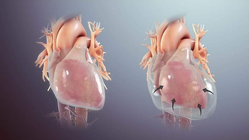
Pericardium
Pericardium
It is the fibro serous sac that encloses the heart and the root of its great blood vessel.
Function
Subdivisions
1. Fibrous pericardium
It is an outer single-layered fibrous sac
2. Serious pericardium
It is an inner double-layer serious sac. Its two-layer are:
1. Parietal pericardium: it is an outer layer
2. Visceral pericardium: It is an inner layer
Difference between the parietal pericardium and serious pericardium
| parietal pericardium | visceral pericardium |
| it is attached to the fibrous pericardium | it is attached to the myocardium of the heart |
| It develops from the somatopleuric mesoderm | it develops from the splanchnopleuric mesoderm |
| it is introverted by somatic nerve fiber | It is innervated by the autonomic nerve fibers |
| It is pain sensitive | It is pain insensitive |
Contents of the pericardium
Arterial supply
Both fibrous pericardium and the parietal layer of the visceral pericardium are supplied by branches of the following artery:
The visceral layer of the serous pericardium is supplied by the coronary arteries.
Nerve supply
Sinuses of pericardium
Between the parietal layer and visceral layers of the serous pericardium, there are two sinuses
1. Transverse sinus
it is a transverse recess behind the ascending aorta and pulmonary trunk and in front of the SVC and superior pulmonary veins. It is developed due to the degeneration of the dorsal myocardium.
It is the horizontal passage between the two pericardial tubes. On each side, it communicates with the general pericardial cavity.
2. Oblique sinus
It is a recess of the serous pericardium behind the base of the heart. It is enclosed by a "J-shaped" sheath of the visceral layer of the serous pericardium enclosing six veins. They are two vena cavae and four pulmonary veins. The oblique sinus permits the distension of the left atrium during the return of oxygenated blood in it from the lungs.
Comments (0)