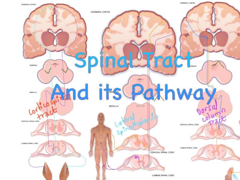
Spinal Tract and Its Pathway

What are nerve fivers?

Axons and dendrites of a neuron (nerve cell processes) are called nerve fibers.  

Tract

Tracts are the bundle of nerve fibers carrying motor & sensory impulses. 

  • Tract forms the majority portion of white matter. 
  • In the spinal cord travel through a white column and synapse with nuclei present in the anterior, lateral, and posterior horn of grey matter of the spinal cord. 


Types of tract

  • Ascending tract (carries the sensory impulse to CNS) 
  • Descending tract (carries motor impulses from the brain) 


Descending (Motor) Tract 

I) Pyramidal (corticospinal tract)  
From the cerebral cortex (a grey matter of the cerebrum) to the spinal cord 
  1. Anterior corticospinal tract — most of the fibers crossed at the corresponding spinal segment 
  2. Lateral corticospinal tract --  most of the fibers crossed at the pyramid of the medulla

II) Extra-pyramidal Tract


 Lateral corticospinal tract

  • 80% of the corticospinal tract crosses the midline — at the level of the medulla (Pyramidal decussation)  
    -- to supply motor function to the opposite side  
  • And descend through the lateral white column of the opposite side of the spinal cord, and supply the opposite side 
    But some of these fibers do not cross the midline and descend through the lateral white column of the spinal cord of the same side as the uncrossed lateral corticospinal tract and supply the same side.

Anterior corticospinal tract 

  • The remaining 20% doesn’t cross the midline at the level of the medulla and descend through an anterior white column on the same side of the spinal cord. 
  • But remember, most of these fibers cross to the opposite side at the corresponding spinal segment to supply the opposite side
Note: Cerebrum is related to contralateral side supply. So each and every tract(Both ascending & descending) arises/terminate from/to the cerebrum, most of them cross the midline and supply to the opposite side. 


Extrapyramidal tract  
From different nuclei present in the brain stem to the spinal cord
 
1) Rubrospinal tract 
From red nucleus to spinal nerve 
 
2) Reticulospinal tract 
From reticular formation To spinal nerve
 
3) Vestibulospinal tract  
From vestibular nuclei to spinal nerve 
 
4) Olivospinal tract 
From the inferior Olivary nucleus to the spinal nerve 

5) Tectospinal tract 

6)Tectobulbar tract (synapse with cranial nerve nuclei) 

Note: Mnemonic RRVOTT 


Descending tract summary Table


The function of the Extra Pyramidal Tract 

1) Rubrospinal tract — Responsible for tone & posture 

2) Vestibulospinal tract — Responsible  for equilibrium  

3) Tecto-spinal tract — Responsible  for Visio-spinal reflex 

4) Coordinate the voluntary moment initiated by the pyramidal tract 

5) When the pyramidal tract is damaged, they may carry the voluntary impulses to some extent 


Pyramidal or corticospinal tract 

Arise from the axon of pyramidal cells present in the cerebral cortex (cerebrum)  

  • 30% from the Primary motor area (area no.4)  
  • 30% from the premotor area (area no.6) 
  • 40 % of the somatosensory area (area no.3,1,2) present in the postcentral gyrus  

                 | 
Fibers pass through corona radiata to the posterior limb of the internal capsule 
                 | 
Pass to crus cerebri of the midbrain and to the Basilar part of the pons  
                |

To pyramid of the medulla oblongata 

The corticospinal tract grouped together to form a swelling known as a pyramid 
Hence, named as pyramidal tract 

            ||  

  1. At the lower part of the pyramid of the medulla, about 80% of the corticospinal tract crosses the midline to go to the opposite side. This is known as pyramidal decussation. These crossed fibers descend in the lateral white column of the spinal cord as the lateral corticospinal tract    

Which terminates by synapsing with anterior horn nuclei, supplying to the opposite side 

      2. About 20% of the tract doesn’t cross the midline and descends in the anterior white column of the spinal cord of the same side as the anterior corticospinal tract

Most of them terminate by crossing the midline and synapsing with opposite anterior horn nuclei, but some end in the same side anterior horn nuclei. 


The function of the pyramidal tract 

  1. Control voluntary movements of the opposite side of the body 
  2. May carry pathways for some superficial reflexes like a cremasteric, abdominal reflex 

Effect of lesion 

  1. Loss of voluntary movements of the opposite half of the body (Spastic type of paralysis) 
  2. Increase muscle tone (muscle tension) 
  3. Reflexes  
  • Superficial reflexes are lost (as the pyramidal tract forms a pathway of superficial reflexes) 
  • Deep reflexes are exaggerated ( as the deep reflex center is within the spinal segment; not in the brain) 
  • Babinski sign is positive  
  • Ankle clonus present  


Upper Motor Neuron And Lower Motor Neuron  

Upper motor neuron 

  • Pyramidal cells and their axon form descending tract within CNS (Brain & spinal cord) 
    Eg. Pyramidal tract & Extrapyramidal tract 

These upper motor neuron synapse in the anterior horn nuclei of the spinal cord giving rise to a spinal nerve, which is termed a lower motor neuron. 

Similarly, upper motor neuron synapses in the cranial nuclei present in the brain stem give rise to the cranial nerve. 


Lower motor Neuron 

Anterior horn nuclei, cranial nuclei, and axon (as spinal & cranial nerves) are known as lower motor neurons. 


Difference Among Upper Motor Neuron & Lower motor Neuron Lesions 

Upper Motor Neuron Lesion Lower Motor Neuron Lesion
Spastic type of paralysis (paralyzed muscles are rigid)
Due to increased muscle tone 

Flaccid type of paralysis
Due to decreased muscle tone 

Deep reflex exaggerated Deep reflex diminished
Babinski sign positive Babinski sign negative
Ankle clonus present Ankle clonus absent
Regeneration absent due to absent in neurolemmal sheath (CNS) Regeneration present due to the presence of the neurolemmal sheath(PNS) 
Muscle wasting absent Muscle wasting present
Lead to contralateral paralysis (as most of the fibers decussated to opposite side)  This leads to ipsilateral paralysis 

Note: But superficial reflex is lost in both 


Difference between the pyramid and extrapyramidal tract 

Pyramidal Tract Extrapyramidal Tract
The anterior and lateral corticospinal tract are called the pyramidal tract All descending tracts except corticospinal tracts are called extrapyramidal tract
Arise from pyramidal cells of the cerebral cortex  Arise from nuclei mainly present in the brain stem
Myelination starts at birth & completed by 2/3 years Myelination started before birth
Rate of conduction slower The rate of conduction is faster
The main function is to control the voluntary movement of the opposite half of the body The main function is to control complex movements of body & tone, posture, and equilibrium


 ASCENDING (Sensory ) Tract 

I) Tract ascending from Posterior white column of spinal cord 
 (These tracts form a Dorsal column-medial lemniscal system) 

 -As passes through the posterior white column in the spinal cord. 
 -In the brain stem passes through the medial lemniscus 

   1. Tract of Gall (Fasciculus Gracillis) 

Carry sensory impulses from the lower half of the body 

   2. Tract of Burdach (Fasciculus cuneatus)  
Carry sensory impulses from the upper half of the body 

II) Tract ascending through the anterior white column of the spinal cord 

Anterior spinothalamic tract 

III) Tract ascending through the posterior white column of the spinal cord 

  1. Lateral spinothalamic tract 
  2. Ventral spinocerebellar tract 
  3. Dorsal spinocerebellar tract 
  4. Spino-vestibular tract 
  5. Spino-olivary tract 
  6. Spino-tectal tract 
  7. Spino-cortical tract 
  8. Spino-reticular tract 
  9. Spino-pontine tract 

Note: mnemonic; Thalamic+cerebellum and VOT CR P 


Table of Important Ascending Tract  

Things to remember  

For every ascending tract— three orders of neurons 

  • 1st order neuron - form from the axon of cells of dorsal root ganglia 
  • 2nd order neuron 1st order neuron synapse with the posterior horn nuclei of the grey matter of the spinal cord (of the same side) 

This means 2nd order neurons arise from the corresponding spinal segment (posterior horn)  

Except for the tract of gall, & tract of Burdach — these tracts don’t synapse in the corresponding grey matter of the spinal segment. They ascend through the posterior (dorsal ) white column of the spinal cord of the same side and synapse with nuclei present in the medulla from where their 2nd order neuron arises

  • 3rd order neuron — arise from nuclei of the thalamus  

 2nd-order neuron synapse with nuclei of the thalamus, giving rise to 3rd-order neuron 

Remember
1st order neuron always related to the same side of the origin
But 2nd order neuron always cross the midline

In case of spinothalamic tract, 2nd order neuron cross at the corresponding spinal segment
In case of Tract of Gall & Burdach, 2nd order neuron arise from nuclei of the medulla and cross the midline at the level of the medulla as internal arcuate fibers (forming sensory decussation)
Note: Cerebrum is related to contralateral side supply. So each and every tract(Both ascending & descending) arise/terminate from/to the cerebrum, most of them cross the midline and supply to the opposite side. 


Tract of Gall (Funiculus Gracillis) and Tract of Burdach (Funiculus Cuneatus) 
[Dorsal column-medial lemniscal system) 

1st order neuron: 

  • Arise from cells of dorsal root ganglia 
  • Enter the posterior white column of the spinal cord of the same side and ascends without synapsing in the nuclei of the posterior horn of grey matter 
  • Then at the medulla synapse with nucleus Gracillis & with nucleus Cuneatus 

2nd order Neuron: 

Arise from axon from nucleus Gracillis & Nucleus cuneatus 
        | 
Its internal arcuate fibers cross the midline and go to the opposite side at the level of the medulla (Forming sensory decussation) 
        | 
In the brain stem, pass through the medial lemniscus 
       | 
Terminate in the thalamus by synapsing with the ventral posterolateral nucleus  
 

3rd order neuron: 

Arise as an axon from the ventral posterolateral nucleus of the thalamus  
       | 
Posterior limb of the internal capsule and end in the somatosensory area (area no.3,1,2) of the postcentral gyrus [cerebral cortex] 


Sensation from the upper part of the body — by Tract of Burdach 

Sensation from the lower part of the body — by Tract of Gracillis 


These tracts carry impulse from the opposite half for: 

  • Fine touch sensation; crude touch by anterior spinothalamic tract 
  • Tactile localization  
  • Tactile discrimination  
  • Kinesthetic sensation (movement) 
  • Vibration sense 
  • Make sensory pathway for superficial reflex 


Spinothalamic tract 

  • Lateral spinothalamic tract  
  • Anterior spinothalamic tract 

1st order neuron: 

  • Arise from the axon of cells of dorsal root ganglia 
  • Then enter in the posterior horn of grey matter of the same side and synapse with its nuclei giving rise to 2nd order neuron 

2nd order neuron: 

Arise from the axon of the nucleus centrodorsalis of the posterior horn    
       
And cross the midline and goes to the anterior white column (ascends as an anterior spinothalamic tract)     

 and lateral white column (ascends as a lateral spinothalamic tract). In spinal cord  
      |  
On reaching the brain stem, lateral and anterior spinothalamic tracts together ascend as the Spinal lemniscus 
      | 
Terminate by synapsing with the ventral posterolateral nucleus of the thalamus from where 3rd order neuron arises  


3rd order neuron: 

This fiber passes through the internal capsule and terminates in the somatosensory area (area no.3,1,2) of the postcentral gyrus. 


Function  

Lateral spinothalamic tract 
— carries pain and temperature sensation from the opposite half of the body 

Anterior spinothalamic tract 
— carries crude touch and pressure sensation from the opposite half of the body 

Tips:  
Exteroceptive sensation: Pain, Temperature, Pressure, Touch sense (PTPT) 
Proprioceptive sensation: Sense of movements, the position of joints, muscle contraction
 
Lemniscus:
Specific tracts which ascend through the white column of spinal cords, on reaching the brain stem, they combine and from a bundle of tracts (appearance like ribbon structure) known as lemniscus. 
  • Medial lemniscus formed by the combination of internal arcuate fibers comes from the opposite side of the nucleus Gracillis & nucleus cuneatus.
  • The spinal lemniscus is formed by a combination of the anterior and lateral spinothalamic tract at the brain stem.

    Other Lemniscus
  • Trigeminal Lemniscus
  • Lateral Lemniscus
 

As a whole

Sensation enters the spinal cord via dorsal roots & ascends in the dorsal white column as a medial lemniscal system and in the anterior and lateral (anterolateral column) as spinothalamic pathways.
The sensory system decussates but at different levels.
  • The medial lemniscal system decussates in the medulla oblongata (forming sensory decussation)
  • While spinothalamic decussate at the level of the corresponding spinal cord.
Motor fibers start from the motor area of the brain,
pass through the corona radiata, 
the posterior limb of the internal capsule & brainstem
In the lowest part of the medulla oblongata, most of the fibers cross to the opposite side and terminate in anterior horn cells of grey matter
Most of the anterior corticospinal tract decussates at the corresponding spinal segment.

Questions collection: Click here