Examination of the Abdomen
- Inspection
- Palpation
- Percussion
- Auscultation
A. Inspection:
Inspect the patient’s abdomen from above, side end, and foot/head end.
On inspection, we see the following things:
1. The shape of the abdomen (Normal, Distended, Sunken)
2. Umbilicus (Inverted, Everted, Slit-like)
3. Flanks (Full/ Not Full)
4. Any visible peristalsis
(We inspect the Site, Direction, and pattern from the side end)
5. Any visible pulsation
6. Engorged veins
We inspect superior venacaval obstruction, inferior venacaval obstruction, and portal HTN by direction of blood flow. i.e.
- In SVCO – blood flow is directed inferiorly,
- IVCO - blood flow directed superiorly, and
- PHTN- blood drains away from the umbilicus)
7. Any abdominal scar marks
8. Skin pigmentation
- (Cullen sign – slightly bluish discoloration in and around the umbilicus,
- Grey turner’s sign – skin discoloration of flanks)
9. Movement of the abdomen with respiration
10. Hernia orifices are intact or not by cough impulse
11. Hair distribution
Presentation of Inspection Finding:
On inspection shape of the abdomen is normal, the umbilicus is centrally placed and inverted, the flanks are not full, no visible peristalsis, no visible pulsation, no engorged veins, no any visible scar marks, no any skin pigmentation, normal hair distribution, hernial orifices are intact.
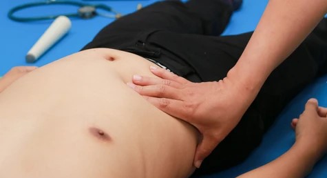
Palpation:
- Superficial palpation
- Deep palpation
- Organ palpation
- Fluid thrill
Note: Before palpating the patient, the hand should be rubbed to make it warm and sanitized properly. Ask the patient whether he/she has pain in any part of the abdomen. If yes then palpate that area at last. Palpation should be started from the left iliac fossa in an anticlockwise direction and end palpation in the umbilicus.
1. Superficial palpation:
We see
- Tenderness,
- Temperature,
- Any lump,
- Muscle guard and Rigidity
(Muscle guard is voluntary resistance during palpation provoked by pain in a painful area whereas rigidity is involuntary muscle contraction)
2. Deep palpation:
We see
- Muscle guard and rigidity,
- Tenderness
(If present then rebound tenderness is done),
- Any masses
(If present then site, size, border, shape, surface, tenderness, consistency, and movement with respiration are noted.)
How to differentiate muscle guard & muscle rigidity?
Ask the patient to relax themselves. If still rigidity is present, ask the patient to semi-flex the hip & knee and check. Still rigidity present, then it is muscle rigidity.
How to deep palpate in case of an ascites patient & in case of an obese patient?
In an obese - one can use both hands
In ascites - Slowly double tap the hand (1st tap to shift the fluid & 2nd tap for actual palpating)
Presentation: On palpation, local temperature is not raised, and the abdomen is non-tender with no palpable mass. The liver is not palpable, spleen and kidney are also not palpable.
3. Organ palpation
I) Liver palpation:
- Find lateral border of rectus abdominis muscle by head rise/leg rise test.
- Then ask the patient to turn the patient’s head towards the left side, the mouth open, and ask the patient to breathe deeply throughout your palpation.
- Now place your right hand on the right iliac fossa (middle finger at lateral border of rectus abdominis and palm parallel to right costal margin).
- Now proceed towards the Right costal margin, 1 cm in every step, and feel the liver margin during deep inspiration. Keep palpating towards the right costal margin and then towards the left lobe side
Measure distance: From the right costal margin along the right midclavicular line up to the palpable liver margin and from the xiphisternum along the midline up to the palpable mass.
Assess - border, surface, consistency, tenderness, thrill.
Upper border of liver dullness:
Note: Usually, no need to percuss the upper border of liver dullness in case of no palpable liver in abdomen examination. (Upper border of liver dullness is checked in the percussion section of respiratory examination)
Start percussion from the right 2nd ICS towards the right lower chest until dullness appears. If dullness appears then locate the area by keeping your right index finger. Start counting the number of ICS where dullness is found from the right 2nd ICS by using your left finger.
Total liver span: Measured from the upper border of liver dullness to palpable liver margin in cm along the right midclavicular line.
MURPHY’S SIGN: Procedure:
- Place your left thumb just below the tip of the 9th costal cartilage (a point that intersects between the transpyloric line and the right midclavicular line).
- Now ask the patient to take a deep inspiration.
- Now press your left thumb firmly during the patient maximum inspiratory phase.
- Note any sudden arrest of breath at the zenith of inspiration.
- Murphy’s sign positive (Present in acute cholecystitis)
Presentation of liver palpation:
- The liver is enlarged x cm from the right costal margin along the right midclavicular line.
- Liver is enlarged x cm from the xiphisternum along the midline.
- Surface is smooth/ irregular.
- Margin is sharp/ rounded/ irregular.
- Liver is tender or not.
- Soft/ firm/ hard in consistency.
- Total liver is x cm
- Upper border of liver dullness is in the right x intercostal space
- Hepatic bruit present or not.
II) Spleen palpation:
Procedure:
- Place your right hand on the patient's right iliac fossa
- Proceed palpation from right iliac fossa towards left hypochondriac region diagonally (move your hand forward during expiration and 1 cm at a time)
- Keep palpating until the splenic margin or left costal margin is felt.
- If the splenic margin is not felt then right lateral decubitus positioning of the patient (left hip and knee semi flexed that resting on patient’s right leg)
- Place your left hand behind the lower left chest (9,10,11th ribs) and pull the rib cage forward and again palpate from the umbilicus up to the left costal & along its margin
Note: If spleen margin is felt then the following are noted-
- Border – regular/irregular
- Surface – smooth/irregular
- Consistency –soft/firm/hard
- Tenderness – tender/non-tender
- Splenic notch – present/ absent
Measurement of the spleen:
From the anterior axillary line up to the palpable tip (towards the right iliac fossa along its long axis)
Or
From the left costal margin where the left midclavicular line intersects (towards right iliac fossa along its long axis)
Presentation to examiner
- The spleen is x cm from the left costal margin towards the right iliac fossa.
- Border – regular/irregular
- Surface – smooth/irregular
- Consistency –soft/firm/hard
- Tenderness – tender/non-tender
- Splenic notch – present/ absent
- Finger insinuation – possible/not possible
III) Kidney palpation:
Right kidney
Bimanual examination
- Place your left hand behind the patient’s right flanks.
- Place your right hand in front of the patient’s right flank
- Now ask the patient to take a deep inspiration
- Now press the fingers of both hands together during the patient’s maximum inspiratory phase.
Ballottement:
- Try to push the patient’s right kidney upwards by your left hand (towards the right hand) and downward by your right hand (towards your left hand)
- Try to feel any bouncing movement of the kidney between your two hands
- If yes kidney is kidney is ballotable
Left kidney
Bimanual examination
- Place your left hand behind the patient’s left flanks.
- Place your right hand in front of the patient's left flank
- Now ask the patient to take a deep inspiration
- Now press the fingers of both hands together during the patient’s maximum inspiratory phase.
Ballottement:
- Try to push patient's left kidney upwards by your left hand (towards the right hand) and downward by your right hand (towards your left hand)
- Try to feel any bouncing movement of the kidney between your two hands
- If yes kidney is kidney is ballotable
Note: Record individually for both right and left kidney
- Bimanually palpable/ not palpable
- If palpable assess size, surface, border, consistency
- Ballotable/not Ballotable
Differentiation of enlarged spleen from enlarged left kidney:
- Mass is Ballotable in the left kidney but not in the enlarged spleen
- Splenic notch is present but absent in the left kidney.
- Finger insinuation is not possible in the enlarged spleen but is possible in the left kidney.
- During percussion dullness in enlarged spleen but resonant in the enlarged kidney (kidney is retroperitoneal, and above it lies intestine which is tympanic)
- In enlarged spleen Movement towards right iliac fossa during respiration but in enlarged left kidney downward movements.
IV) Urinary bladder examination
Palpation: Place your ulnar border on left just above the umbilicus. Palpate vertically downward up to pubic symphysis. Or by using both hands make a downward V-shaped
Percussion: Start percussion in the midline from the area of resonance towards pubic symphysis. Stop percussion where dullness appears.
If any swelling felts assess it's:
- Shape – oval/pear
- Surface – smooth/irregular
- Margin – definite/indefinite
- Consistency – firm/cystic/hard
- Side-to-side movement – possible/not possible
How to differentiate ovarian cyst or uterus or urinary bladder?
V) Aorta and para-aortic lymph nodes examination;
Palpation: Place the tips of both the right and left fingers of the hand to the left of the umbilicus and push deeply. Now go for deep palpation and feel the abdominal aorta and just lateral to that, try to palpate the lymph nodes and move to upward direction palpating in the same manner.
Note: Any palpable round, firm fixed para-aortic lymph nodes.
C. Percussion:
1. Shifting dullness;
- Start percussion from below the xiphisternum in the midline.
- Continue percussion up to the umbilicus & go to the right or left flanks until dullness appears.
- Stop percussion where resonance changes into dullness in the flanks.
- Ask patients to roll towards his/her left side and wait for at least 10 sec
- Again start percussion from the point of dullness towards the midline.
- If dullness changes into resonance again, shifting dullness is positive. (Positive in case of ascites)
2. Fluid thrill; Procedure
- Ask patients to place his/her ulnar border of the left hand in the midline of the abdomen.
- Now place your left hand to the left side of the abdomen.
- Now flick the right side of the abdomen by any of your right fingers.
Note: Try to feel any ripple in your hand. If ripple felt, fluid thrill positive
D. Auscultation
- Bowel sound: To the right and below the umbilicus
(To declare bowel sound absent - one should auscultate up to 3 minutes)
- Aortic bruits: Just to the left and above the umbilicus.
- Renal bruits: 2-3 cm above from the midline of the umbilicus and then 2-3 cm lateral to it. (On both sides)
- Liver bruits: Over the liver
- Splenic rubs: Over the spleen
Comments (0)