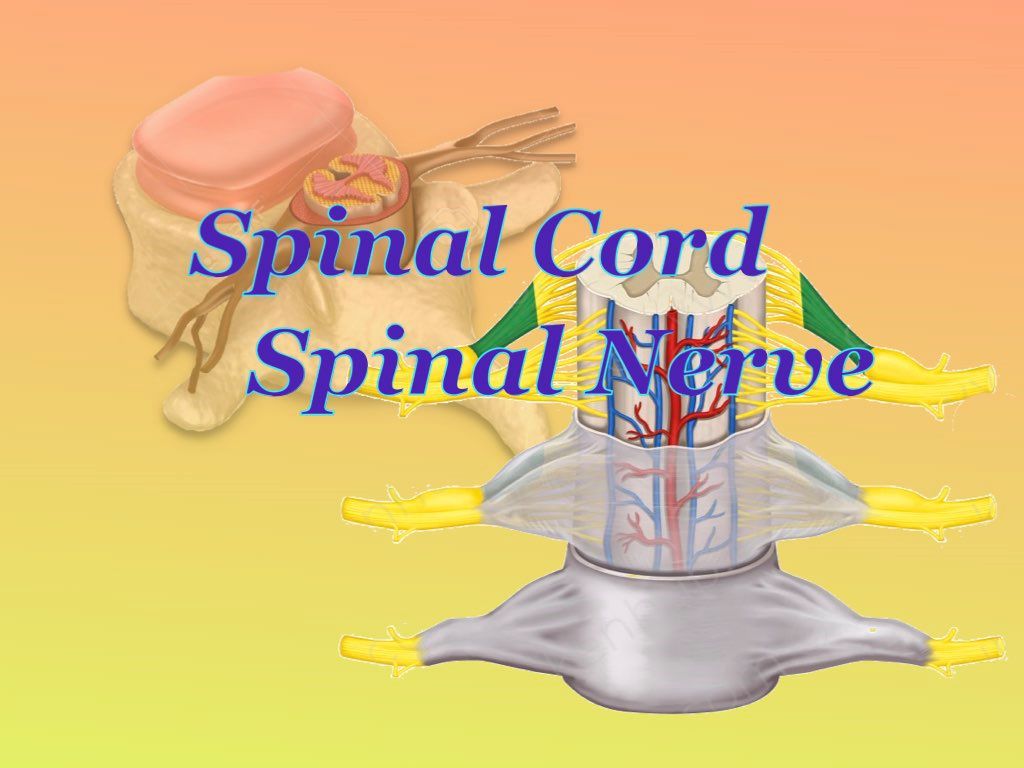
Spinal nerve
Formed by the union of the ventral root & dorsal root of the spinal cord within the intervertebral foramen.
Ventral root carry fibers come from anterior horn nuclei(motor in function) and arise from the anterolateral sulcus of the spinal cord
Dorsal root carry fibers come from posterior horn nuclei of grey matter (sensory in function) that arise from the postero-lateral sulcus of the spinal cord
| 31 pairs of Spinal nerve |
33 vertebrae |
| 8 cervical |
7 cervical vertebrae |
| 12 thoracic spinal nerve |
12 thoracic vertebrae |
| 5 lumber nerve |
5 lumbar vertebrae |
| 5 sacral |
5 sacral vertebrae fused as one |
| 1 coccygeal nerve |
4 coccygeal vertebrae fused as one |
All spinal nerve passes through the intervertebral foramen except:
-
1st cervical nerve — passes through the dorsal part of the superior articular surface of the C1 vertebra
- 5th sacral nerve and 1st coccygeal nerve through sacral hiatus
Difference between ventral root and dorsal root of spinal nerve
| Ventral Root |
Dorsal Root |
|
No ganglion in the ventral root
|
Dorsal root ganglia (contain pseudounipolar neuron).
1st-order neurons of all ascending tracts arise from this
|
| Ventral root motor in function |
Dorsal root sensory function |
Ganglia are the collection of the nerve cell body in PNS (spinal nerve and spinal cord)
Nuclei are the collection of nerve cell body within the CNS (brain and spinal cord)
Eg. Basal nuclei, red nucleus, cranial nuclei, spinal nuclei (anterior horn and posterior horn nuclei)
Branches of spinal nerve
Dorsal Ramus — supplies dorsal body wall (not to limbs)
Ventral Ramus — supplies ventral body wall and also upper limb and lower limb.
Functional components of spinal nerve
Main theme: Spinal nerve is a mixed nerve
- Sensory from body wall & viscera
- Motor to muscle & viscera
General somatic afferent(GSA) sensory from the body wall to dorsal root ganglia
General visceral afferent(GVA) sensory from viscera to dorsal root ganglia
General somatic efferent(GSE) motor to skeletal muscle
General visceral efferent(GVE) motor to viscera(smooth and cardiac muscle)
Content of vertebral canal (formed by combined vertebral foramen)
- Spinal cord with its meninges and blood vessels ie. one anterior and two posterior spinal arteries
Spinal meninges ie. dura mater, (arachnoid mater, pia mater together form leptomeninges)
Potential space between meninges
- Epidural space between spinal dura mater & periosteum of the vertebral canal
- Subdural space (below dura mater)
- Sub Arachnoid space (below the arachnoid, between the pia and arachnoid mater) contains CSF and blood vessels
Note: Spinal dura mater only consists of the meninges layer of dura mater.
This means spinal dura mater is the continuation of the meningeal layer of cranial dura mater
Spinal leptomeninges is also the continuation of the cranial leptomeninges
The cranial dura mater contains both the endosteal and meningeal layers of the dura mater.
Intervertebral foramen (formed by inferior vertebral peduncle & superior vertebral peduncle)
Here, the Formation of spinal nerves occur
Spinal cord
Length: 45cm
Extension: From the medulla at the level of the upper border of the atlas to the lower border of the 1st lumbar vertebra.
In the case of the child, up to L3 vertebra
Superiorly continues as medulla oblongata and inferiorly terminates as conus medullaris.
- The lower tapering end of the spinal cord is called conus medullaris.
- The extension of the pia mater below the level of conus medullaris is called the film terminal.
- Dorsal and ventral nerve roots of L2-L5, S1-S5 & Co1 are collectively known as cauda equina.
I) Dura mater and arachnoid mater extend up to the lower border S2 vertebra.
II) Subarachnoid space (between the arachnoid mater and the pia mater) also extends up to the S2 vertebra.
Thus CSF only presents up to the S2 vertebra.
But the pia mater extends up to 1st segment of the coccyx.
Below the conus medullaris, the pia mater gets fused and extends as a thread-like structure called filum terminalis.
- Below the lower border of L1, the spinal cord is absent. And up to S2 CSF present. That’s why lumbar puncture can be done between below L2to S2
- Actually done between L3-L4vertebra or L4-L5
- Lumbar puncture is a procedure to withdraw CSF( colorless fluid) for various examinations.
Filum terminals:
The Pia mater surrounding the spinal cord gets fused below the conus medullaris and extended as thread-like structures.
Up to the dorsal of 1st piece of coccyx
Two parts:
1) Filum terminalis internum:
As Duramater & arachnoid mater extended up to the sacral segment, filum terminals within this sheath are called filum terminals internal.
2) Filum terminalis externum:
Below the 2nd sacral segment, no dura & pia mater covering the Filum terminalis
Extend up to 1st coccygeal segment
Transverse section of Different spinal segment
Cervical segment
- Cervical enlargement to supply upper limb (origin of brachial plexus ie. root value C5- T1)
- No lateral horn
- The anterior horn (motor in function) is broader than the posterior horn as the motor supply to the upper limb.
Note: Ventral ramus which arises from the anterior horn of the grey matter of the spinal cord only takes part to form the plexus.
The dorsal ramus doesn’t form the plexus.
Thoracic segment
- No enlargement
- Anterior horn and posterior horn narrow; no supply to the limb
- Lateral horn present.
Lateral horn only presents between T1-T12 and L1 & L2 segment for the supply of viscera. Form part of the autonomic nervous system; present for thoracolumbar flow.
Give rise to preganglionic sympathetic fibers.
Sympathetic fibers from the lateral horn — the ventral root of the spinal nerve — Trunk of the spinal nerve — Ventral ramus — sympathetic ganglion (collectively form sympathetic chain)
The lateral horn also present in Sacral S2-S4, forms pelvic splanchnic nerves for visceral supply ie. hindgut, urinary bladder, genital organs, sexual function (erection= complex vascular and parasympathetic process)
Autonomic nervous system (a kind of motor supply)
Sympathetic nerve: Thoracolumbar outflow
Parasympathetic nerve: Craniosacral outflow (III, VII, IX, X Cranial nerve)
Lumbar segment
- Lumbar enlargement due to origin of lumbar plexus( for lower limb supply)
- Lateral horn only present in L1& L2
- The anterior & posterior horn is broad.
Sacral segment
- No enlargement
- Lateral horn present (S2-S4) for pelvic splanchnic nerves
Features of the spinal cord
Anterior median fissure lodge for anterior spinal artery, formed by the fusion of branches of 4th part of (Rt. & Lf.)vertebral arteries
Two posterolateral sulci each lodge for posterior spinal artery, the branch of 4th part of vertebral artery
Internal structure
- Outer white matter
- Inner grey matter
- The center of grey matter has central canal containing CSF
But in the cerebrum & cerebellum, Outer grey matter & inner white matter.
In the brain stem(midbrain, pons, medulla) —outer white matter with dispersed nuclei(as grey matter ) within the white matter.
Division
- Each half is divided into anterior, lateral & posterior region by anterolateral & posterolateral sulcui.
- Ventral motor root arises from anterolateral sulcus.
- The dorsal sensory root arises from the posterolateral sulcus
Grey matter
- Anterior horn- consists of motor nuclei
The axon of these nuclei give lies at the ventral root of the spinal nerve
- Posterior horn- consists of sensory nuclei
The axon of these sensory nuclei gives rise to the dorsal root of the spinal nerve
- The lateral horn is present only in T1-L2 which gives rise to sympathetic fibers for visceral supply which also passes through the ventral root of the spinal nerve
And lateral horn in S2-S4 gives arise to parasympathetic fibers with also pass through the ventral root
Note: There are no other parasympathetic fibers arising from the spinal cord rather than S2-S3.
All other parasympathetic fibers are from the cranial nerve(3,7,9,10)
Division of white matter
- Anterior white column (Anterior funiculus)
- Lateral White column (Posterior funiculus)
- Posterior white column(Lateral funiculus)
Note: Tracts (collections of nerve fibers) travel through these white columns
- Descending tract(motor) travels through the white column and only synapses with nuclei of anterior horn nuclei of grey matter for supply.
- Similarly ascending tracts (sensory) after synapse with posterior horn nuclei of grey matter, ascend and travel through these white columns.
Note: Collectively, nerve fibers form tracts in CNS (brain & spinal cord) and this nerve fiber after synapse with nuclei of grey matter emerges as PNS( cranial nerve & spinal nerve)
And tracts form a major max. portion of white matter.
White matter is called a pathway of nerve fibers(tracts)
White matter consists of
- Nerve fibers, blood vessels & neuroglia
Grey matter consists of
- Nerve cell body,
- Blood vessels and neuroglia (supporting cells)
Note: Collection of nerve cell body form nuclei/ nucleus in CNS
Eg.anterior, lateral & posterior horn nuclei of the grey matter of the spinal cord
In the brain— basal ganglia, thalamus, intracerebellar nuclei, red nucleus, Edinger-Westphal nucleus
Collection of the nerve cell body in PNS( spinal & cranial nerve) form ganglia.
Eg.dorsal root ganglia in the spinal nerve, 4 parasympathetic ganglia in the cranial nerve, sympathetic chain/ ganglia in the ventral ramus of T1-L2 spinal nerves
Commissure means the connection between two hemispheres
- Connection Between white matter of two hemispheres — White commissure
- Connection Between grey matter of two hemispheres— Grey commissure
Nuclei present in the posterior horn of grey matter of the spinal cord
- Posteromarginal nucleus
- Substantial gelatinosa—For pain pathway
- Nucleus proprius
- Nucleus dorsalis
Blood supply of the spinal cord
Artery supply
- One Anterior spinal artery (lodge in anteromedian fissure)
Formed by the fusion of branches from the 4th part of the right and left vertebral arteries.
_Supply anterior 2/3rd of spinal cord
- Two posterior spinal arteries (lodge in posterolateral sulci)
Branches of 4th part of vertebral artery
_Supply posterior 1/3rd of spinal cord
Venous drainage
To six longitudinal channels
- One anteromedian vein
- One posteromedian vein
- Two Anterolateral vein
- Two posterolateral vein
These all drain to the intervertebral venous plexus
Histology of spinal cord
Embryology/ development of spinal cord
Develop from neural tube (neuroectoderm)
The neural tube — is formed by folding and closure of the neural plate.
During neural tube formation, cells outside the neural tube form neural crest cells, which migrate to different places and form
All ganglia
- Cranial nerve ganglia
- Parasympathetic ganglia (3,7,9,10)
- Dorsal root ganglia of spinal nerve
- Sympathetic chain/ganglia of T1-L2 segment of spinal nerve
Comments (0)