Kidney
5 years ago 3086
Location of the kidney
Either side of the vertebral column, from T12 to L3 vertebrae.
Situated in the left hypochondrium, epigastric and umbilical region.
The right kidney is slightly lower in position due to its relationship with the liver.
Functions of the kidney
| Eliminate nitrogenous waste products from protein metabolism through urine. |
| Maintain water and electrolyte balance. |
| Maintain acid-balance. |
| The kidneys secrete renin which helps in the auto-regulation of blood. |
| The kidneys secrete erythropoietin for the maturation of RBC. |
| The kidneys secrete 1:25hydroxycholecalciferol for the control of calcium metabolism. |
Presenting parts of the kidney
Two Surfaces: Anterior & Posterior
Two Ends: Upper & Lower
Two borders: Medial & Lateral
| Anterior Surface is convex |
| Posterior Surface is flat |
| Medial border is convex-concavo-convex |
| Lateral border is convex |
What factors keep the kidneys in position?
What are the coverings of the kidneys?
From within outwards
Fibrous capsule is formed by condensation of the fibrous stroma of peripheral of the kidney. The capsule covers the entire organ, lines the renal sinus, and is reflected as a tubular sheath around the minor and major calyces and pelvis of the ureter.
Perinephric fat lies between the interval of fibrous capsule and renal fascia
Para nephric fat lies between the interval of renal fascia and the anterior layer of thoraco-lumbar fascia. It is abundant on the Posterior surface.
Renal fascia distribution:
It is formed by condensation of fibro-areolar tissue surrounding the kidneys and perinephric fat.
It has two layers:- anterior and posterior deposition of renal fascia
| Laterally- both layers fuse at the lateral border and are continuous with the fascia transversalis |
| Medially- * Anterior layer covers the anterior surface of the kidneys and renal vessels and is continuous with a similar layer in front of the abdominal aorta and inferior vena cava. * Posterior layer covers the posterior aspect of the kidneys and renal vessels, bends with the psoas muscle fascia, and attached to the bodies of the lumbar vertebra. |
| Above- Both the layers fuse at the upper end of the kidneys and split to enclose the supra-renal gland in a separate compartment. At the upper end of the supra renal gland, they reunite and is continuous with the sub diaphragmatic fascia forming the suspensory ligament of the supra renal gland. |
| Below- They do not fuse and extend downwards along the ureters and finally lost in the extra peritoneal tissue of the iliac fossa |
Relations of the anterior surface of the kidney
It is partially covered with the peritoneum, the relations differ between right and left kidneys
Right kidney:
| Suprarenal area (non-peritoneal) |
| Duodenal area (non-peritoneal) - related to the second part of the duodenum. |
| Hepatic area (peritoneal) - related to the inferior surface of the right lobe of the liver. |
| Colic area (non-peritoneal) - related to right colic flexure. |
| Jejunal area (peritoneal) - related to coils of jejunum. |
Left kidney-
| Suprarenal area (non-peritoneal) |
| Splenic area (peritoneal) - related to the lower part of the visceral surface of the spleen. |
| Gastric area (peritoneal) - related to the posteroinferior surface of the stomach. |
| Pancreatic area (non-peritoneal) - related to the body of pancreas and splenic vessel. |
| Colic area (non-peritoneal) - related to left colic flexure and descending colon. |
| Jejunal area (peritoneal) - related to coils of jejunum. |
Relations of the posterior surface of the kidney
The posterior surface is entirely non-peritoneal.
Upper part:-(from within outwards)
| The diaphragm |
| Medial and lateral arcuate ligament |
| Costo-diaphragmatic recess of pleura |
| 11th & 12th ribs on the left side, 12th ribs on the right side. |
Lower part:-
| Psoas major, quadratus lumborum, and transverses abdominis muscle |
| Para nephric fat |
| Subcostal vessel and nerve |
| Ilio hypo-gastric and ilio-inguinal nerves |
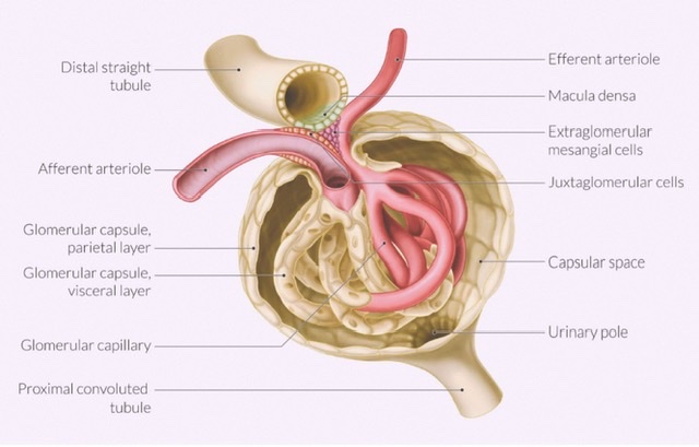
What is hilum? Name its contents.
It is a deep vertical fissure at the central area of the medial border, bounded by anterior and posterior lips. At the level of the L1 vertebra
Contents of Hilum:
| Renal artery (Entering kidney) |
| Renal vein (Exiting kidney) |
| Pelvis of the ureter (Exiting kidney) |
| Lymphatics (exiting kidney) |
| Nerves (entering kidney) |
| Fat |
Structure of the kidney
It can be classified into two ways:
Macroscopic has two parts:-
Renal substance consists of:
Renal cortex consists of:
Renal medulla includes:
Renal sinus is the space within the kidney lined by the renal capsule. It contains:-
Microscopic has two parts:-
What is the pelvis of the ureter?
It is a funnel-shaped dilation of the ureter, formed by the union of major calyces. It extends from the renal sinus through the hilum to the caudal pole of the kidney where it is continuous with the abdominal part of the ureter.
Capacity:- 5-7ml
Artery supply of the kidney
Renal artery, a direct branch of abdominal artery
Venous drainage of the kidney
Each kidney is drained into the inferior vena cava by the corresponding left and right renal vein.
Left renal vein has two tributaries:- left gonadal vein & the left suprarenal vein.
Lymphatic drainage of the kidney
Drain into lateral aortic lymph nodes.
Nerve supply of the kidney
Sympathetic renal plexus arise from the T10-L1 segment of the spinal cord. Sympathetic helps in vaso-motor and stimulates the juxta-medullary cells to release renin.
Parasympathetic renal nerves are derived from both vagus nerves.
Development of the kidney
The matured kidneys are developed from the intermediate mesoderm in the craniocaudal direction. It has three stages:
a) At first, pronephros develops.
b) Pronephros is then replaced by mesonephros.
c)Mesonephros is replaced by metanephros (Permanent kidney}
Metanephros has two parts:-
DEVELOPMENTAL ANOMALIES
1) Lobulated kidney: Results from the persistence of fetal lobulation.
2) Pelvic kidney: Due to failure of the ascent of the kidney.
3) Horse-shoe shaped kidney: Due to non-union of the two metanephric blastema across the midline.
4) Unilateral double kidney: Two kidneys on the same side.
Also read: Anatomy Question Collection
Also read: Anatomy Questions & Answers
Also read: Anatomy notes
Comments (0)