Gall Bladder
5 years ago 3231
Gallbladder is an elongated pear-shaped sac
It is located in the fossa for the Gall bladder on the inferior surface of the right lobe of the liver.
Part of GallBladder
Fundus:
Anteriorly: Anterior Abdominal Wall
Posteriorly: Transverse Colon
Body:
Above: The liver
Below: Transverse colon & 1st part of the duodenum
Right side: Right Colic Flexure & Superior Duodenal flexure
Left side: Pyloric part of Stomach
Neck:
Above: The liver
Below: 1st part of the duodenum
Peritoneal Relations of Gallbladder:
| Fundus: Lower expanded free end (covered with peritoneum on all sides) |
| Body: Extend from the fundus to the neck (cover with peritoneum except the upper surface) |
| Neck: 'S' shaped extends from body to cystic duct( covered with peritoneum except for upper part) |
| Upper surface of the body is completely related to the Liver so it is devoid of peritoneum |
Structure of Gallbladder:
The wall of the Gallbladder consists of 3 coats.
From outside to inside:
The mucosa is lined by simple columnar epithelium
Note: No submucosa is present
Artery supply of Gallbladder:
Cystic artery branch of the hepatic artery.
Venous drainage: of Gallbladder
Cystic vein drain into the portal vein
This small organ has many anomalies As like
Clinical Importance
Amazingly, it is more frequent in 5F’s i.e Fair, Fertile, Fatty Female & above Forty years of age
Nice to know:
Gallbladder is an extrahepatic biliary apparatus.
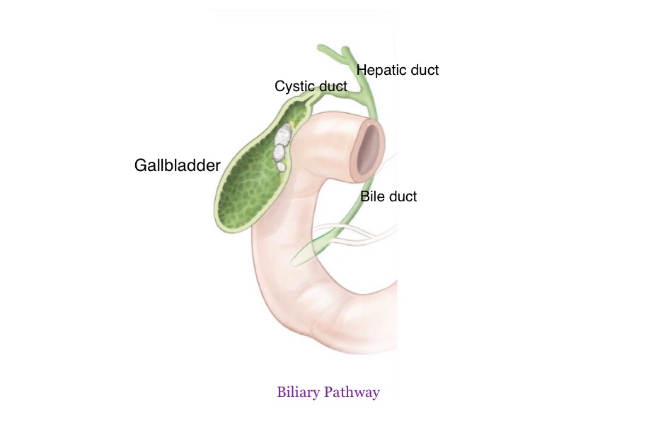
Biliary Apparatus
Biliary apparatus: The biliary or excretory apparatus of the liver includes the passage through which hepatic bile and gall bladder bile are conveyed into the second part of the duodenum.
Parts of Biliary Apparatus:
Parts of intra-hepatic biliary apparatus:
| Bile canaliculi: They form a polygonal network around individual liver cells of the hepatic lobules except those surfaces of the cells which are related to sinusoids. |
| Canal of Hering: They are formed by the union of canaliculi in the peripheral part of hepatic lobules. |
| Bile ductules: They occupy interlobular spaces. |
| Right & left hepatic ducts: They are formed by the union of bile ductules with one another that drain the bile respectively from the physiological right and left lobes of liver. |
Parts of extra-hepatic biliary apparatus:
| Common hepatic duct: It is formed by the union of right & left hepatic ducts close to the right end of porta hepatis. |
| Gall bladder: It is a pear-shaped hollow viscus, situated obliquely in a non-peritoneal fossa on the undersurface of the right lobe of liver, and extends from the right end of porta hepatis to the inferior border of the liver. |
| Cystic duct: It begins from the neck of the gall bladder and ends by joining the right side of the common hepatic duct. |
| Bile duct (common): It is formed by the union of common hepatic and cystic ducts and unites with the main pancreatic duct before termination to form a dilatation known as the ampulla of Vater in the posteromedial wall of the second part of the duodenum. |
Also read: Anatomy Question Collection
Also read: Anatomy Questions & Answers
Also read: Anatomy notes
Comments (0)