Liver
5 years ago 5120
The liver is the largest gland of the body.
Liver color: Red-brown.
Shape of Liver: Wedge-shaped.
Location of Liver:
Liver occupies-
| Whole of the right hypochondrium |
| Greater part of the Epigastrium |
| Left hypochondrium |
Anatomical points of Liver
This is the viscera of the liver with the gall bladder.
| The right lobe is slightly higher than the left lobe. |
| Groove for inferior vena cava lies posteriorly and directed vertically downwards. |
| Fossa for gall bladder lies inferiorly. |
Why liver is called a mixed gland?
Because the liver consists of both exocrine and endocrine parts.
| Exocrine part secretes bile which is drained out through the biliary pathway. |
| Endocrine part liberates some useful chemical substances such as glucose from glycogen, most of the plasma protein(except the immunoglobulins), and heparin directly into the bloodstream. |
Here hepatocytes perform both endocrine & exocrine functions.
Function of Liver:
The liver secretes bile & stores glycogen.
It synthesis the serum proteins & lipids.
It detoxifies blood from endogenous & exogenous substances (e.g., toxins, drugs, alcohol, etc) that enter the circulation.
It produces hemopoietic cells of all types during fetal life.
Why is the liver much larger in children?
Due to hematopoietic function in fetal life, and occupies about 2/5th of the abdomen.
What is the basic tissue of the liver?
Epithelial Tissue
What will happen if we cut down the 2/3rd portion of the liver?
It will regenerate itself because the cells of the liver are stable cell (or Ito cell) which has regeneration capability.
Supports of the liver:
| Hepatic vein opening into inferior vena cava |
| Intra-abdominal pressure |
| Ligaments of the liver |
Ligaments of Liver:
True Ligament of Liver:
Ture ligament is derived from the remnants of the embryological structures.
| Ligament teres hepatis represents the obliterated umbilical vein. |
| Ligamentum venosum represents obliterated ductus venosus. |
False ligament of Liver:
False Ligament is formed from the fold of the peritoneum.
| Falciform ligament |
| Coronary ligament |
| Right Triangular ligament |
| Left triangular ligament |
| Lesser omentum |
External features of the Liver:
The liver presents two surfaces:
But it can be categorized altogether on 5 surfaces.
Border of Liver: Sharp inferior border.
Lobes of Liver:
Anatomical & Physiological lobes
Anatomical Right & Left lobes Division of Liver:
On the diaphragmatic surface:
The liver is divided into 2 right & left lobes by the attachment of the Falciform Ligament.
On the visceral surface:
Liver is divided into right & left lobes by the fissures for ligamentum teres hepatis, ligamentum venosum.
Physiological Right & Left lobes division of Liver:
On the diaphragmatic surface:
The liver is divided into right & left lobes from the line passing from the little right of the attachment of falciform ligament.
On the Postero-inferior surface:
Liver is divided into right & left lobes from the imaginary line passing from the center of the groove of the inferior vena cava and fossa of the gallbladder (Cholecysto-venacaval Line).
Anatomically quadrate & caudate lobe belongs to the right lobe.
Physiologically quadrate & caudate lobe belongs to the left lobe.
| Physiological right lobe | Physiological left lobe |
| supplied by the right branch of hepatic artery | supplied by a left branch of hepatic artery |
| supplied by the right branch of portal vein | supplied by a left branch of portal vein |
| Bile from the right physiological lobe is carried by the right hepatic duct | Bile from left physiological lobe is carried by left hepatic duct |
What is Porta Hepatis?
It is a transverse non-peritoneal fissure & acts as a gateway of the liver.
Structures entering into porta hepatis:
| Right & left branches of the hepatic artery. |
| Right & left divisions of the portal vein. |
| Hepatic plexus of nerve. |
Structures emerging out of porta hepatis :
|
Right & left division of hepatic ducts
|
|
Cystic duct
|
|
Lymph vessels.
|
Remember: Hepatic Veins don't pass through the porta hepatis, thus porta hepatis is not the true gateway.
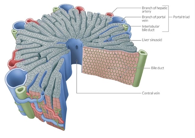
What are the contents of the portal triad?
Hepatic artery, portal vein, and bile ductule
Peritoneal relation of Liver
Most of the area of the liver is covered by the peritoneum. The area of the liver that is not covered by the peritoneum is covered by Glisson's capsule.
The areas which are not covered by the peritoneum are:
| Bare area of the liver |
| Fossa for gall bladder |
| Groove for inferior vena cava |
| Porta hepatis |
| Groove for ligamentum venosum |
| Groove for ligamentum teres hepatis |
| Area of attachment of Falciform ligament |
Bare area of liver
The bare area of the liver (non-peritoneal area) is a large triangular area on the diaphragmatic surface of the liver, devoid of peritoneal covering.
Superior coronary ligament
Inferior coronary ligament
Right triangular ligament
Left triangular ligament
Why bare area of the liver is called "The Bare Area"?
Because it is the largest non-peritoneal area of the liver.
Importance of the bare area
It is devoid of peritoneum so it directly comes into contact with the diaphragm which separates it from the right pleural sac.
Portosystemic anastomosis takes place here.
Why there remain a concavity between two convex parts on the superior surface of the liver?
For Cardiac Impression
What do you mean by portosystemic Anastomosis?
Anastomosis between the tributaries of portal vein and tributaries of the systemic vein.
What are the sites of portosystemic anastomosis?
| Lower end of the esophagus |
| Anorectal Junction |
| Around the umbilicus. |
| Posterior abdominal wall |
| Bare area of liver |
| Attachment of falciform ligament. |
How is the Nutrition of the liver maintained?
66% to 75% from the portal vein.
25% to 33% from the Hepatic artery.
How the inferior vena cava is formed?
By the union of right and left common iliac vein at the level of the 5th lumbar vertebra.
How portal vein is formed?
By the union of superior mesenteric vein and splenic vein behind the neck of the pancreas at the level of Lumbar 2 vertebra.
What is the importance of the right lateral surface of the liver?
Needle biopsy of the liver is approached through the 9th or 10th intercostal spaces at the mid-axillary line during forced expiration in order to avoid injury to the lung.
Biliary Apparatus
Biliary apparatus: The biliary or excretory apparatus of the liver includes the passage through which hepatic bile and gall bladder bile are conveyed into the second part of the duodenum.
Parts of Biliary Apparatus:
Parts of intra-hepatic biliary apparatus:
| Bile canaliculi: They form a polygonal network around individual liver cells of the hepatic lobules except those surfaces of the cells which are related to sinusoids. |
| Canal of Hering: They are formed by the union of canaliculi in the peripheral part of hepatic lobules. |
| Bile ductules: They occupy interlobular spaces. |
| Right & left hepatic ducts: They are formed by the union of bile ductules with one another that drain the bile respectively from the physiological right and left lobes of liver. |
Parts of extra-hepatic biliary apparatus:
| Common hepatic duct: It is formed by the union of right & left hepatic ducts close to the right end of porta hepatis. |
| Gall bladder: It is a pear-shaped hollow viscus, situated obliquely in a non-peritoneal fossa on the undersurface of the right lobe of liver, and extends from the right end of porta hepatis to the inferior border of the liver. |
| Cystic duct: It begins from the neck of the gall bladder and ends by joining the right side of the common hepatic duct. |
| Bile duct (common): It is formed by the union of common hepatic and cystic ducts and unites with the main pancreatic duct before termination to form a dilatation known as the ampulla of Vater in the posteromedial wall of the second part of the duodenum. |
Histological structure of the liver
Hepatic lobule- called a venous unit of liver.
Portal lobule- called a nutritional unit of liver.
Portal acinus- called structural/metabolic unit of liver.
What is hepatic lamina?
When 20 sheet hepatocytes arrange in a radiating manner, it is known as the hepatic lamina.
What is hepatic lacuna?
The space between two hepatic laminae is called hepatic lacuna.
What is the space of Disse?
Between the walls of the sinusoid and hepatic laminae of cells there exist e potential space called space of Disse.
What is the Space of Mall?
Spaces of Mall are the potential spaces that appear in the interlobular spaces between the Glisson's capsule of the portal canal and the hepatic plates of the cell.
What are the events that occur in the sinusoid?
Physiological shunt occurs and hepatic artery & portal vein drains into here.
What are the other side of the sinusoid rather than the liver?
Spleen, bone marrow, adenohypophysis.
Why liver lymph contains more plasma protein?
The lymph which produces in Space of Mall contain higher plasma protein. That's why the liver contains it more.
What is the origin of the hepatic vein? Intrahepatic or Extrahepatic?
Intrahepatic
Development of Liver
Ventral wall of the endoderm of the foregut.
Also read: Anatomy Question Collection
Also read: Anatomy Questions & Answers
Also read: Anatomy notes
Comments (0)