Large Intestine
5 years ago 4113
Length: About 1.5 m long.
Extension: From the caecum in the right iliac fossa to the anus in the perineum.
Lining Epithelium of Large gut
Simple columnar epithelium with intersperse goblet cells
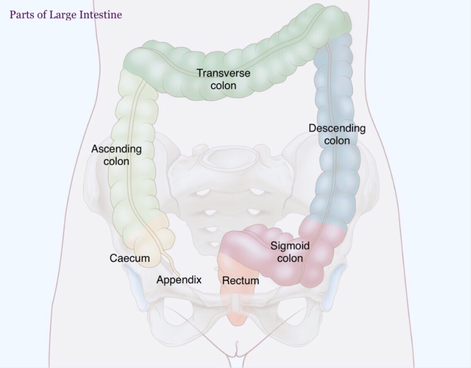
Parts: It has four parts:
The colon is further divided into four parts:
Cardinal Feature of Large Intestine:
a) Teniae coli
These are three ribbon-like bands of the longitudinal muscle coat.
Location: In the caecum, ascending colon & descending colon. Absent in appendix, rectum, & anal canal.
Positions of teniae in ascending colon are
But in the transverse colon, the corresponding positions are inferior, posterior, and superior, respectively.
b) Appendices Epiploicae:
These are small bags of visceral peritoneum filled with fat attached to the teniae of the large intestine. Thus, they are absent in the appendix, rectum, and anal canal.
c) Sacculation (or Haustrations):
These are a series of pouches/dilatations in the wall of the caecum and colon between the teniae.
The sacculations are responsible for the characteristic puckered appearance of the large intestine.
CAECUM
The caecum is the large dilated blind sac at the commencement (proximal end) of the large intestine.
It communicates:
Ascending Colon
The ascending colon is an upward continuation of the caecum.
Extension: From the caecum, at the level of the ileocaecal orifice, to the hepatic (right colic) flexure.
Transverse Colon
The most mobile part of the large intestine.
Extension: From the right colic flexure (in right lumbar region) to the left colic flexure (in the left hypochondriac region).
Descending Colon
Extension: From the left colic flexure to the front of the left external iliac artery at the level of pelvic brim where it becomes continuous with the sigmoid colon.
Sigmoid Colon
Extension: From the lower end of descending colon at the left pelvic inlet to the pelvic surface of the third sacral vertebra where it becomes continuous with the rectum.
Rectum:
It is the continuation of the sigmoid colon at the level of sacral 3 vertebrae.
Length: 12 cm
It has 2 types of curvature
| Antero-posterior curvature 1. Sacral curve 2. Perineal curve |
| Lateral curvature 1. Upper 2. Middle 3. Lower |
Peritoneal relation of Rectum:
| Upper 1/3rd: Covered with peritoneum in front & on both side |
| Middle 1/3rd: Covered only in the front |
| Lower 1/3rd: Devoid of peritoneum |
Visceral relations of Rectum:
Anteriorly in males:
| Rectovesical pouch (upper 2/3rd) |
| Base of the urinary bladder |
| Terminal parts of ureters |
| Seminal vesicles |
| Prostate |
Anteriorly in females
| Rectouterine pouch (Upper 2/3rd) |
| Lower part of the vagina (lower 1/3rd) |
Posteriorly: Same in males and females
| Lower 3 pieces of the sacrum, coccyx |
| Piriformis, Coccygeus, Lecator ani muscles |
| Sympathetic chain |
| Lymph nodes, Lymphatics |
| Fat |
Interior of the rectum:
Two types of fold
| Temporary (longitudinal type, disappear when distended) |
| Permanent: Known as Houston's Valve, 4 in number |
Nerve supply:
Sympathetic: By superior Hypogastric Plexus (L1, L2) vasomotor
Parasympathetic: Pelvic splanchnic nerves, secretomotor to glands
Importance of Rectal vein:
They are the site of portosystemic anastomosis
They're devoid of valves. In portal obstruction, veins became distended and form primary internal piles
Anal canal:
The terminal portion of the large intestine below the pelvic diaphragm
Length: 3.8 cm
Extension: Anorectal junction to the anal orifice
Interior of Anal Canal:
Divided into upper, intermediate, and lower parts by a pectinate line & Hilton's line respectively
1. Upper part: 1.5 cm in length
| Area above the pectinate line |
| Lined by Simple columnar epithelium |
| Internal rectal venous plexus lies outside, gives the "plum red" appearance |
| Feature: Anal columns Anal valves Anal papillae Anal Sinuses Anal glands |
2. Intermediate Part
| Area between Pectinate to Hilton's line |
| Lined by Non-Keratinized stratified squamous epithelium, |
| Internal rectal venous plexus gives a bluish pink appearance. |
3. Lower Part (8 mm)
| Area below Hilton's Line |
| Lined by true skin (Keratinized stratified squamous epithelium) |
| Sweat, sebaceous gland presents |
Nerve supply :
| Above the pectinate line: (like rectum) Sympathetic: Parasympathetic: |
| Below the pectinate line: Somatic spinal nerve via inferior rectal nerves |
Somatic means pain, temperature, touch pressure sensation occur here.
Difference between Male & Female Anal Canal
Male has hair follicle in the lower part of the anal canal but in female no hair follicle.
Anal Fissure:
When hard, large feces passage during bowel movement, then a small tear may occur in the mucosa of the anal canal resulting in a painful condition called Anal Fissure.
Infected glands act as a source of anal fissure.
What is a hemorrhoid (Piles)?
Dilatation, elongation, and tortuosity of anorectal venous plexus formed by radicals of superior middle and inferior rectal veins. In portal obstruction of superior rectal veins, veins are distended and form primary internal hemorrhoids
Types:
| Internal or true piles: Piles that appear above the pectinate line. Painless, bright red blood, |
| External or false piles: Piles that appear below the pectinate line. Very painful, don't bleed. |
Peritoneal Relations of Large Gut:
All the parts of the large intestine are retroperitoneal & fixed except the appendix, transverse colon & sigmoid colon which are intraperitoneal and possess mesenteries.
| Part of Large gut | Name of mesentery |
| Appendix | Mesoappendix |
| Transerve colon | Transverse mesocolon |
| Sigmoid colon | Sigmoid mesocolon |
Functions of the large intestine
| Absorption of water from fluid contents in it to help form the feces. |
| Storage, lubrication, and expulsion of feces. |
| Synthesis of vitamin B complex by normal bacterial flora presents its lumen. |
| Protection from invasion by microorganisms by its mucoid secretion which is rich in IgA group of antibodies. |
Difference between Right colic Flexure & Left colic Flexure
| Right colic Flexure | Left colic Flexure |
| Also called Hepatic flexure | Also called Splenic Flexure |
| Located in the Right lumber region | Located in the Left hypochondrium region |
| Lining epithelium developed from the endoderm of the midgut | Lining epithelium developed from the endoderm of the hindgut |
Difference between the Rectum with the rest of the Large Intestine.
Cardinal features of Large Intestine like Teniae coli, Appendices Epiploicae, Sacculation is absent in Appendix.
Also read: Anatomy Question Collection
Also read: Anatomy Questions & Answers
Also read: Anatomy notes
Comments (0)