Stomach
5 years ago 5487
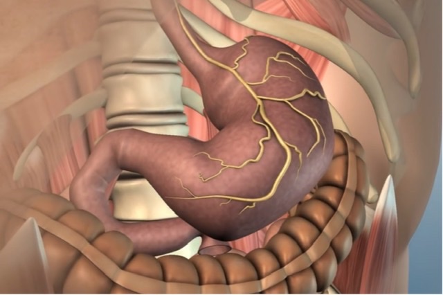
Stomach is the widest and most distensible part of the alimentary canal between the esophagus and the duodenum.
Situated in the left hypochondriac, umbilical, and epigastric regions. It extends obliquely from the left hypochondriac region into the epigastric region.
Shape: 'J ' shaped muscular bag
Size and Capacity
Length: 25 cm
| Organ having the same length as stomach (25 cm) |
| Duodenum, esophagus, descending thoracic aorta, ureter. |
Capacity: In adults, the capacity is 1500 to 2000ml
Function of stomach
| Form a reservoir of food. |
| Mixes food with gastric secretions to form a semi-fluid substance called chyme. |
| Controls the rate of delivery of chyme into the small intestine to allow proper digestion and absorption in the small intestine. |
| Hydrochloric acid secreted by the gastric glands destroys bacteria present in the food and drink. |
| Castle's intrinsic factor present in the gastric juice helps in the absorption of vitamins in the small intestine. |
EXTERNAL FEATURES
| Two ends: Cardiac and Pyloric |
| Two curvatures: Greater and Lesser |
| Two surfaces: Anterior (anterosuperior) and Posterior(posteroinferior) |
How to distinguish between cardiac orifice & pyloric orifice?
| Cardiac orifice is a wider opening & the muscular layer is thin. |
| Pyloric orifice is a narrow opening & is usually thicker due to the presence of a pyloric sphincter. |
Vertebral level of cardiac end and pyloric end
Cardiac end: T11
Pyloric end: L1
Lesser Curvature
| Form the right border of the stomach. |
| Provides attachment to the lesser omentum. |
| Contents of lesser Omentum: Anastomosis of right and left gastric vessels. |
Greater Curvature
| Forms the longer left border of the stomach. |
| Provides attachment to the greater omentum, gastrosplenic, and gastrophrenic ligaments. |
| Contents of Greater Omentum: Anastomosis of Right and Left Gastroepiploic vessels. |
Surfaces
Anterosuperior surface: Faces forward and upward.
Posteroinferior surface: Faces backward and downward.
Parts of Stomach
| 1. Cardiac part: Present cardiac orifice |
| 2. Fundus: Upper dome-shaped part of the stomach situated above the horizontal plane drawn at the level of the cardiac notch. Superiorly the fundus usually reaches the level of the 5th intercostal space just below the nipple, hence gastric pain sometimes imitates the pain of angina pectoris. |
| 3. Body The body and pyloric part are divisible into two parts by the sulcus intermedius. Pyloric antrum(7.5cm) and pyloric canal(2.5cm). |
| 4. Pyloric part: A line is drawn downward and to the left from an angular notch to the greater curvature separates it from the body. It is divided into three parts: - Pyloric antrum - Pyloric canal - Pylorus |
Content of Fundus of the stomach
Fundic Gas (50ml)
Difference between the fundus of the stomach and the fundus of the uterus?
| Fundus of the stomach has a cavity whereas the Fundus of the uterus doesn't have any cavity. |
| Fundus of the stomach contains fundic gas but the fundus of the uterus doesn't contain any gas. |
How pyloric sphincter form?
The circular muscle of the stomach thickened to form a pyloric sphincter.
The sphincter of the cardiac end is called the physiological sphincter and the pyloric sphincter is called the anatomical sphincter.
RELATIONS
Peritoneal Relations
The stomach is covered by the peritoneal except
| Along the Greater & Lesser Curvature, where the blood vessels run. |
| Small area posteriorly near the cardiac orifice. |
The peritoneal folds extending from the lesser and greater curvature of the stomach to other structures are as following
1. Lesser omentum
2. Greater omentum
3. Gastroplenic ligament
4. Gastrophrenic ligament
Why greater omentum is called "Abdominal Policeman"?
Because greater omentum Wraps an inflamed organ and prevents the spread of infection to the general peritoneal cavity.
Visceral Relations
1. Relations of the anterior surface:
| On the right side, related to the Gastric impression of the left lobe of the liver |
| Near the pylorus to the quadrate lobe liver. |
| On the left side, related to the diaphragm and rib cage. |
| The lower part of this surface is related to the anterior abdominal wall. |
2. Relations of the posterior surface:
(Structures Forming Stomach Bed)
| Diaphragm |
| Transverse mesocolon |
| Left colic flexure |
| Splenic artery |
| Pancreas |
| Spleen |
| Left kidney |
| Left suprarenal gland |
MICROSCOPIC STRUCTURE
Consists of four coats/ histological layers.
From the outside inward,
Serous,
Muscular,
Submucous,
Mucous coats
Layers of gastric mucosa
Glands are always present in Lamina Propria except Brunner's gland of Duodenum, which is present at sub-mucosa.
Lamina propria contains three types of gland
Lining epithelium of the stomach
Simple Columnar epithelium (From abdominal part of the esophagus to pectinate part of the anal canal)
[Throughout the GI tract except for esophagus and lower part of anal canal lining epithelium - Simple columnar epithelium]
Exception: Thoracic part of the esophagus and lower part of anal canal lining epithelium lined by Non keratinized stratified squamous epithelium
INTERIOR OF THE STOMACH
When the stomach is cut open, the interior of the stomach presents the following features.
| Gastric folds: The mucosa of an empty stomach is thrown into numerous folds called gastric rugae. |
| Gastric pits: These are small depression on the mucosal in which opens the gastric glands. |
| Gastric canal: A longitudinal furrow that forms temporarily during swallowing between the longitudinal folds of the mucosa along the lesser curvature. |
How gastric canal is formed?
It is produced due to the contraction of the right free margins of the oblique muscle.
ARTERY SUPPLY
| Left gastric artery, a direct branch from the coeliac trunk. |
| Right gastric artery, a branch of the common hepatic artery. |
| Left gastroepiploic artery, a branch of the splenic artery. |
| Right gastroepiploic artery, a branch of the gastroduodenal artery. |
| Short gastric arteries, branches of the splenic artery. |
Examples of Tortuous Arteries
| Facial artery |
| Splenic artery |
| Uterine Artery |
| Nutrient Artery |
[Mnemonics: FUNS]
What is the importance of a splenic artery as a tortuous artery?
It allows the distention of the stomach and helps in well circulation of blood.
Why stomach is supplied by the branches of the coeliac trunk?
The coeliac trunk is the artery of the foregut and stomach is a derivative of the foregut.
VENOUS DRAINAGE
| Left gastric vein |
| Right gastric vein |
| Left gastroepiploic vein |
| Right gastroepiploic vein |
| Short gastric veins |
NERVE SUPPLY
Sympathetic Innervation
The sympathetic fibers are derived from T6 to T10 spinal segments via greater splanchnic nerves, and coeliac and hepatic plexuses.
The sympathetic supply to the stomach is
a) vasomotor
b) motor to the pyloric sphincter, and inhibitory to the remaining gastric musculature, and
c) serves as the Chief pathway for pain sensations from the stomach.
Parasympathetic Innervation
The parasympathetic fibers are derived directly from the vegas nerves.
CLINICAL ANATOMY
Gastric carcinoma:
Commonly occurs along the greater curvature of the stomach as the maximum of the lymphatic vessels passes through this area to drain into pancreatico-splenic lymph node.
Gastric Ulcer:
Commonly occurs along the lesser curvature due to the presence of gastric canal through which liquid substance of food flows directly along with it.
Importance of Gastric Triangle:
If an esophageal obstruction occurs, a small tube passes through the gastric triangle for feeding to the patient.
Also read: Anatomy Question Collection
Also read: Anatomy Questions & Answers
Also read: Anatomy notes
Comments (0)