General Histology 2
6 years ago 23560
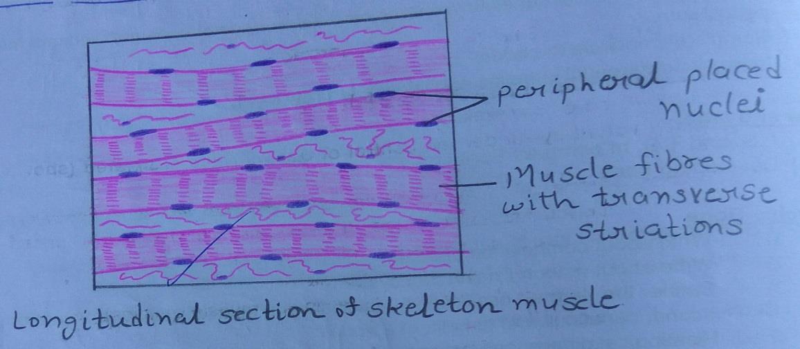
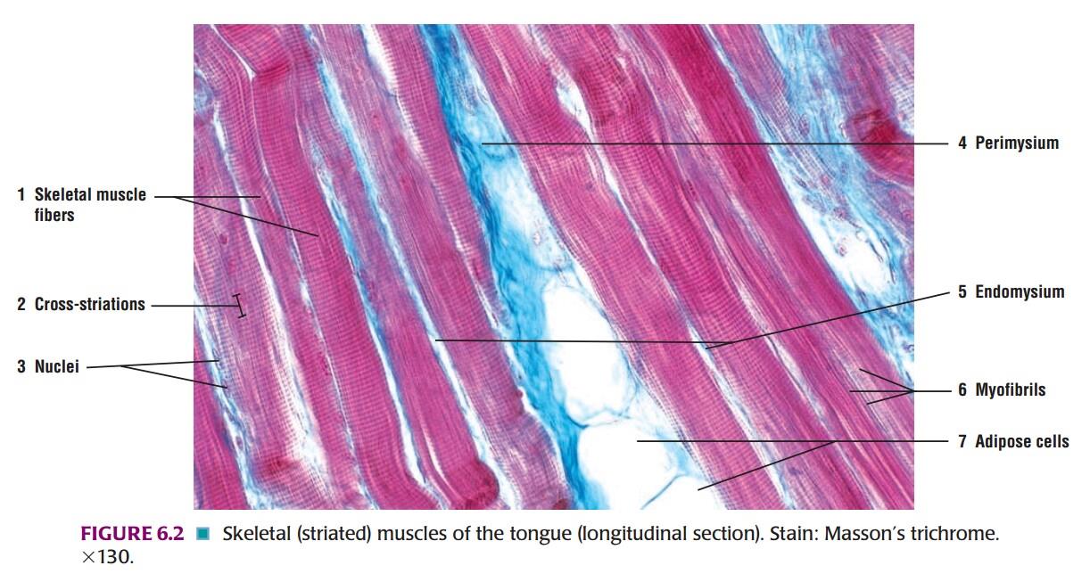
Slide 1: Longitudinal Section of SKELETON MUSCLE
DRAWING:
IDEAL SLIDE:
IDENTIFICATION POINTS:
| 1. In a longitudinal section through the skeleton muscle, the fibers are easily distinguished as they show characteristic transverse striations. |
| 2. The fibers are long and parallel without branching. |
| 3. Many flat nuclei are placed at the periphery. |
| 4. The muscle fibers are separated by some connective tissue. |
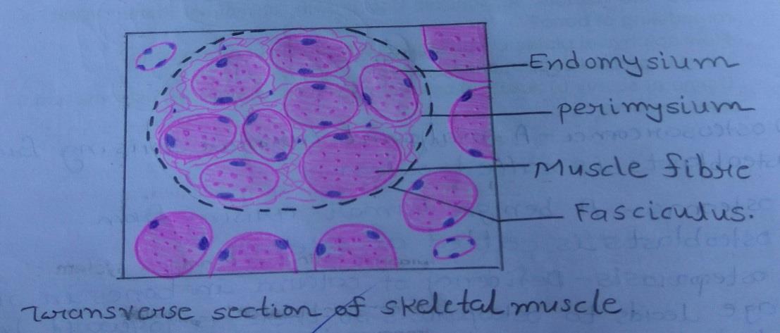
Slide 2: Transverse Section of SKELETON MUSCLE
DRAWING:
IDEAL SLIDE:
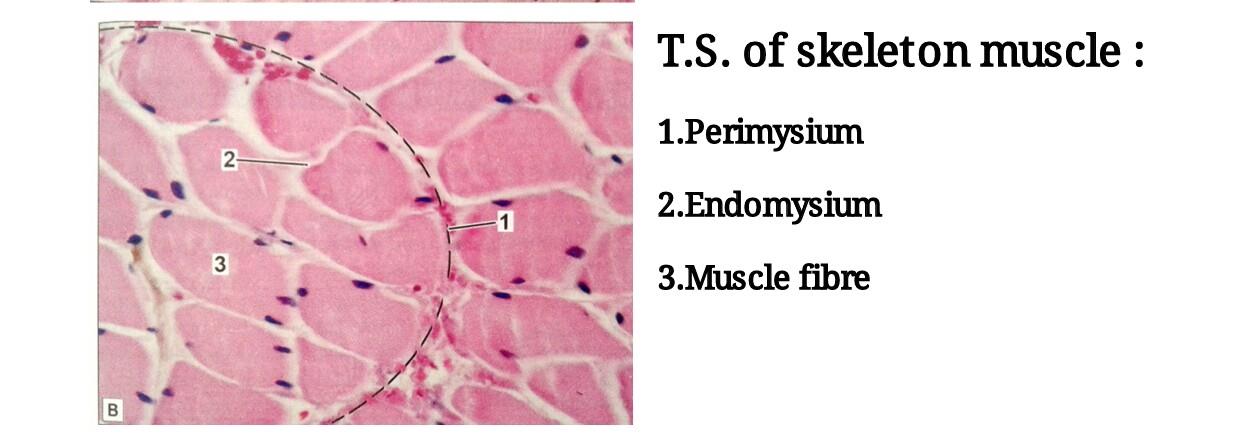
IDENTIFICATION POINTS:
| 1. Fibres seen as irregularly round structures with peripheral nuclei. |
| 2. Muscle fibers grouped into numerous fasciculi. |
| 3. Dots within the fibers are myofibrils which are seen at higher magnification. |
| 4. The connective tissue of the muscle consists of: - Epimysium: connective tissue sheath of muscle. - Perimysium: connective tissue covering of each fascicle. - Endomysium: loose connective tissue surrounding each muscle fiber. |
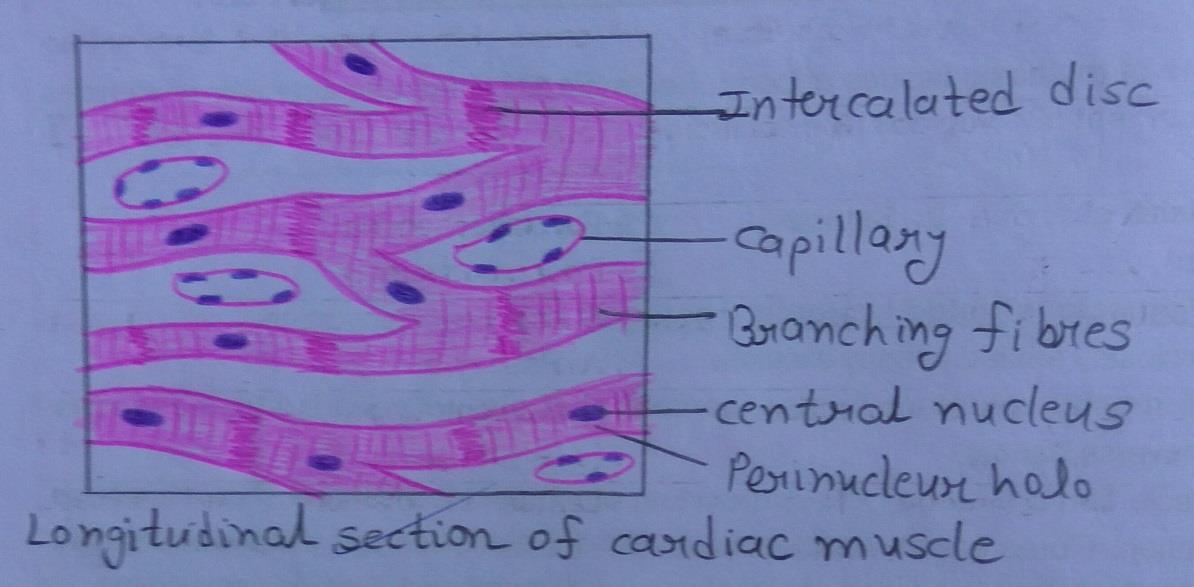
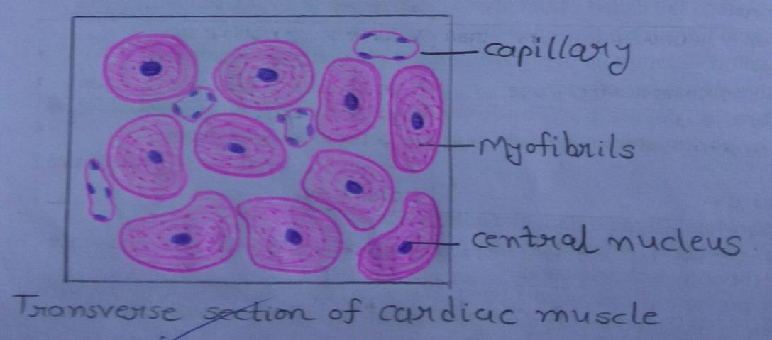
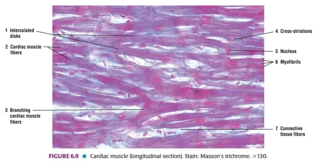
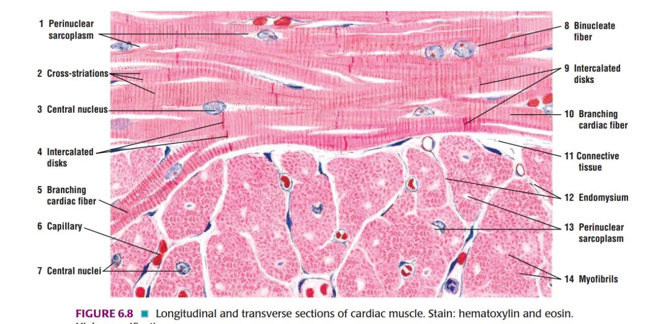
Slide 3: CARDIAC MUSCLE
DRAWING:
IDEAL SLIDE:
IDENTIFICATION POINTS:
| The fibers of cardiac muscle do not run in strict parallel formation, but branch and anastomose with other fibers to form a network. |
| Each fiber of cardiac muscle is not a multinucleated syncytium as in skeleton muscle but is a chain of cardiac muscle cells each having its own nucleus. |
| The nucleus of each myocyte is located centrally and not peripherally as in the skeleton muscle. |
| The myofibrils and striations of cardiac muscle are not as distinct as those of skeleton muscle. |
| With the light microscope, the junctions between adjoining cardiac myocytes are seen as dark staining transverse lines running across the fibers, but are broken into a number of ‘steps. |
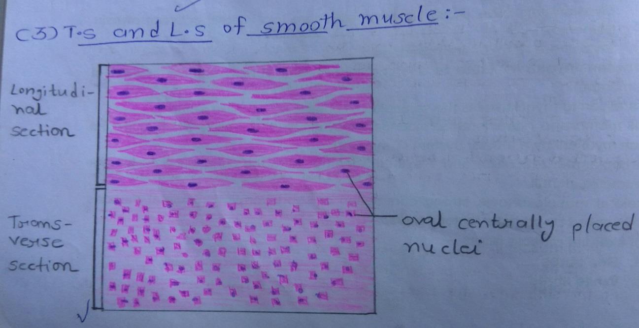
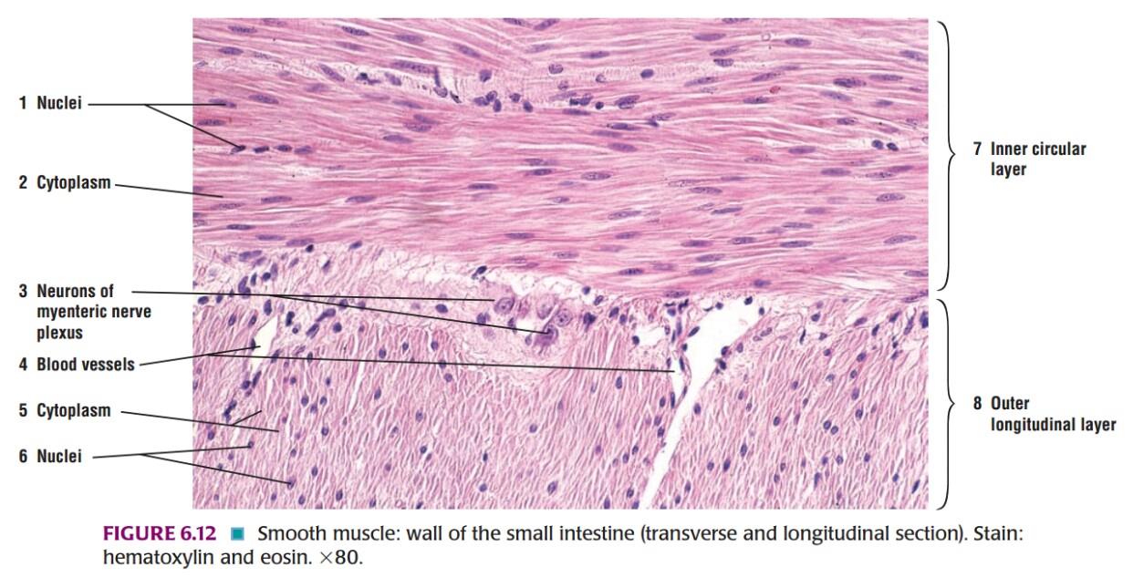
Slide 4: SMOOTH MUSCLE
DRAWING:
IDEAL SLIDE:
IDENTIFICATION POINTS:
| 1. It is a fusiform/spindle-shaped cells having a broad central part and tapering ends. |
| 2. The nucleus, which is oval or elongated, lies in the central part of the cell. |
| 3. With the light microscope, the sarcoplasm appears to have indistinct longitudinal striations but there are no transverse striations. |
| 4. In such a layer, the cells are so arranged that the thick central part of one cell is opposite the thin tapering ends of adjoining cells. |
| 5. In the longitudinal section, the nucleus is elongated and centrally placed. |
| 6. In the transverse section, the nucleus is seen in those cells which are cut through the center but others do not show nuclei. |
Nervous Tissue Histology
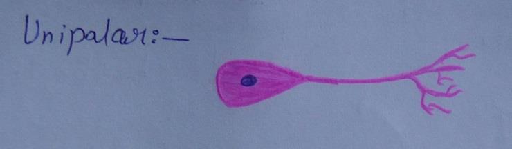
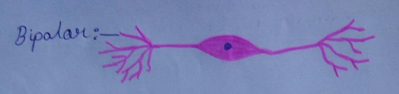
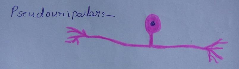
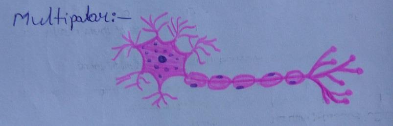
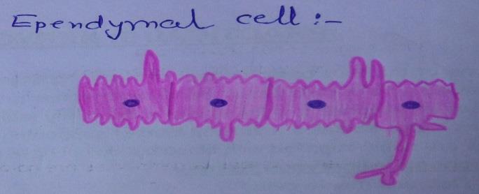
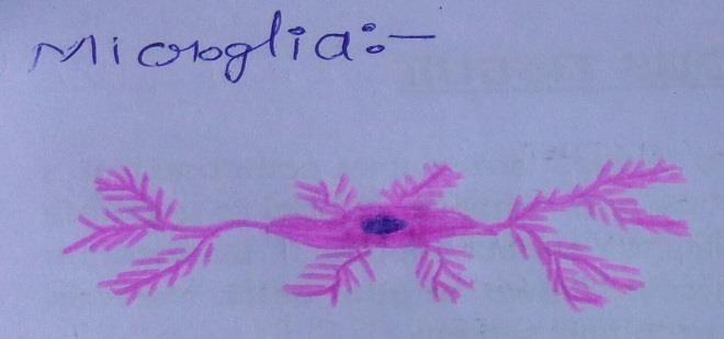
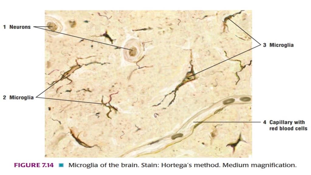
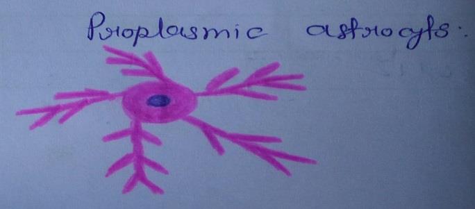
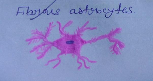
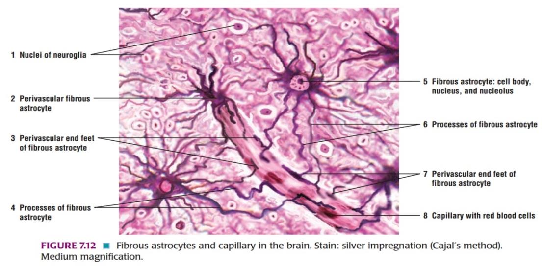
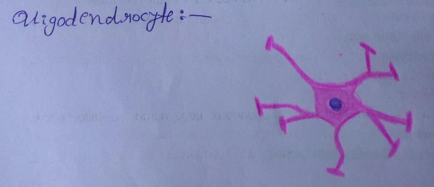
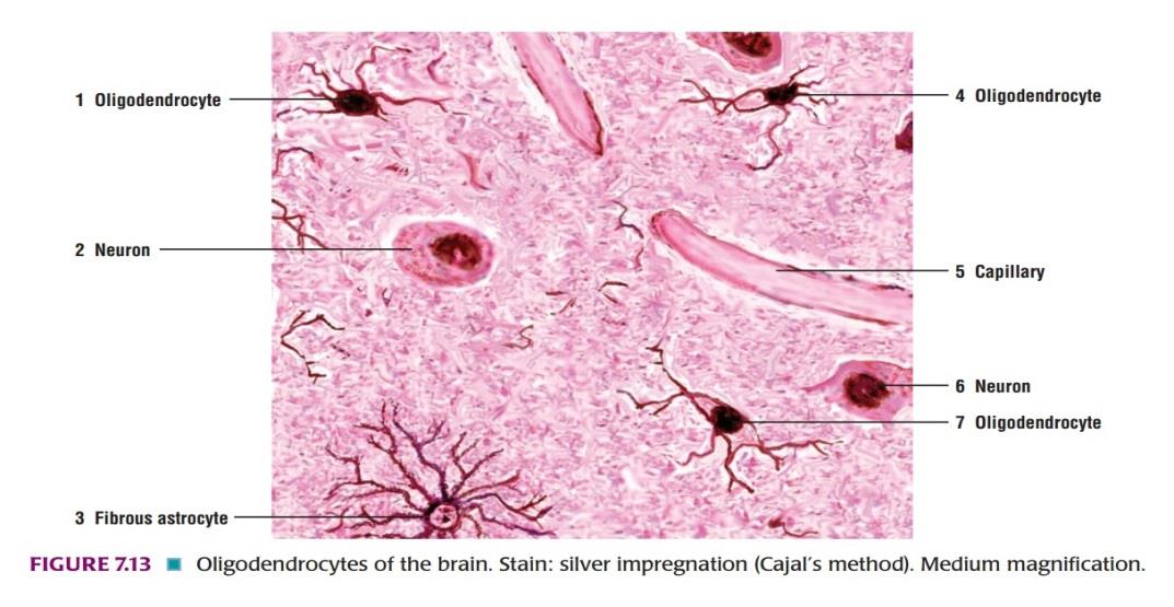
Slide 5: DIFFERENT TYPES OF NEURONS AND NEUROGLIA CELLS
TYPES OF NEURONS:
TYPES OF NEUROGLIA CELLS:
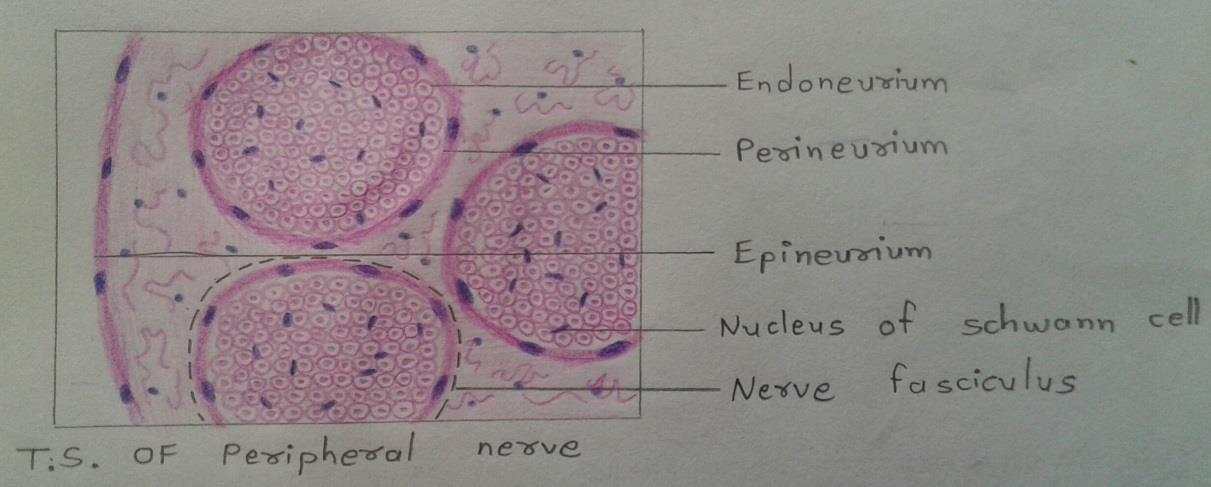
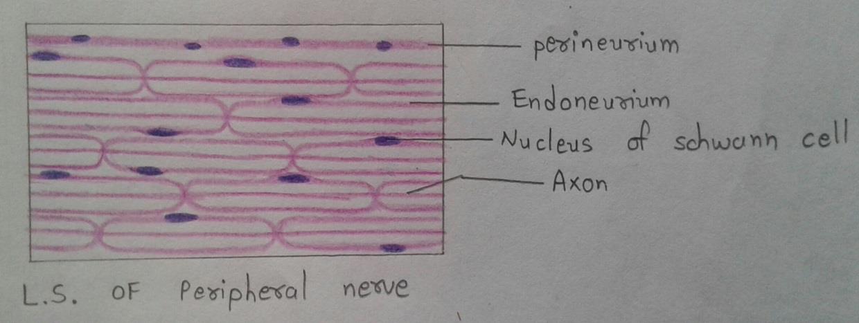
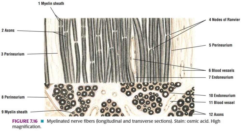
Slide 6: PERIPHERAL NERVE
DRAWING: (H & E STAINING)
IDEAL SLIDE:
IDENTIFICATION POINTS:
| 1. In a longitudinal section of the peripheral nerve, the central axons appear as slender threads stained lightly with H & E staining. |
| 2. In a longitudinal section, the individual axons usually follow a characteristic wavy pattern. |
| 3. Located among the wavy axons in the nerve fascicle are numerous nuclei of the Schwann cells and fibrocytes of the endoneurium. |
| 4. However, it is often difficult to distinguish between the nuclei of Schwann cells and the fibrocytes of the endoneurium. |
| 5. In a transverse section, the axons appear as thin, dark central structures, surrounded by the dissolved remnants of myelin. |
Lymphatic System Histology
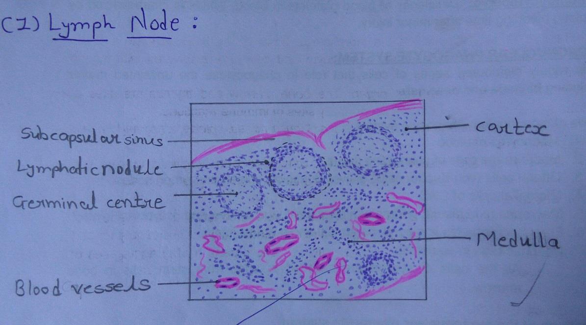
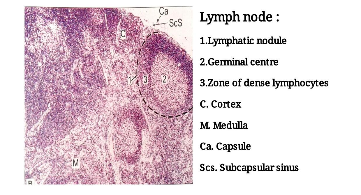
Slide 7: LYMPH NODE
DRAWING:
IDEAL SLIDE:
IDENTIFICATION POINTS:
| 1. A thin capsule surrounds the lymph node and sends in trabeculae. |
| 2. Just beneath the capsule a clear space is seen. This is the subcapsular sinus. |
| 3. A lymph node has an outer cortex and an inner medulla. |
| 4. The cortex is packed with lymphocytes. A number of rounded lymphatic follicles are present. |
| 5. Each nodule has a pale staining germinal center surrounded by a zone of densely packed lymphocytes. |
| 6. Within the medulla the lymphocytes are arranged in the form of anastomosing cords. |
| 7. Several blood vessels can be seen in the medulla. |
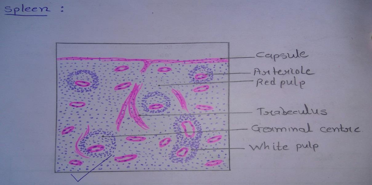
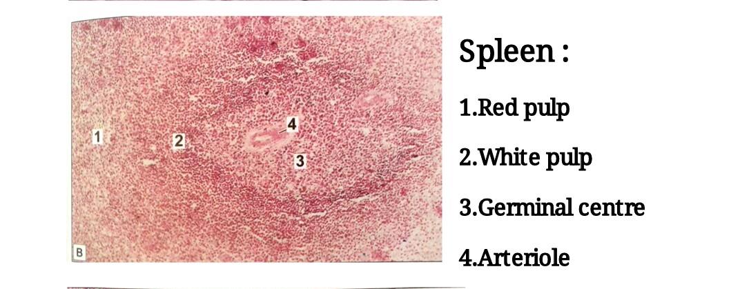
Slide 8: SPLEEN
DRAWING:
IDEAL SLIDE:
IDENTIFICATION POINTS:
| The spleen is characterized by a thick capsule with trabeculae extending from it into the organ. |
| The substance of the organ is divisible into the red pulp in which there are diffusely distributed lymphocytes and numerous sinusoids and the white pulp in which dense aggregation of lymphocytes are present. The latter are in the form of cords surrounding arterioles. |
| When cut transversely, the cords resemble the lymphatic nodules of lymph nodes and like them, they have germinal centers surrounded by rings of densely packed lymphocytes. |
| The nodules of the spleen are easily distinguished from those of lymph nodes because of the presence of arterioles in each nodule. |
| This arteriole occupies an eccentric position in the nodules. |
| More than one arteriole may be present in relation to one germinal center. |
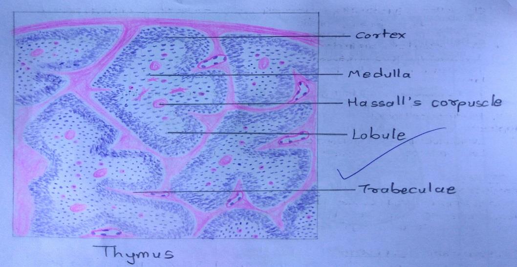
Slide 9: THYMUS
DRAWING:
IDEAL SLIDE:
IDENTIFICATION POINTS:
| 1. The thymus is made up of lymphoid tissue arranged in the form of distinct lobules. |
| 2. The presence of this lobulation enables easy distinction of the thymus from all other lymphoid organs. |
| 3. The lobules are partially separated from each other by connective tissue septae. |
| 4. In each lobule an outer darkly stained cortex and an inner lightly stained medulla are present. |
| 5. Whereas the cortex is confined to one lobule, the medulla is continuous from one lobule to another. |
| 6. The medulla contains pink staining rounded masses called the corpuscles of Hassall. |
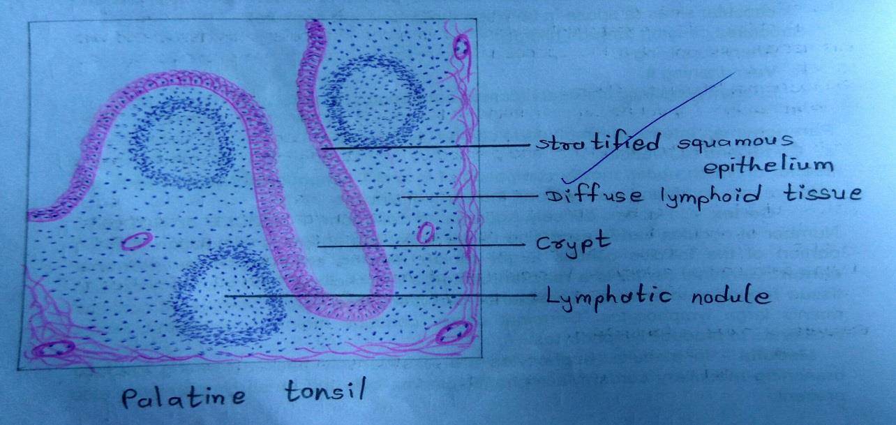
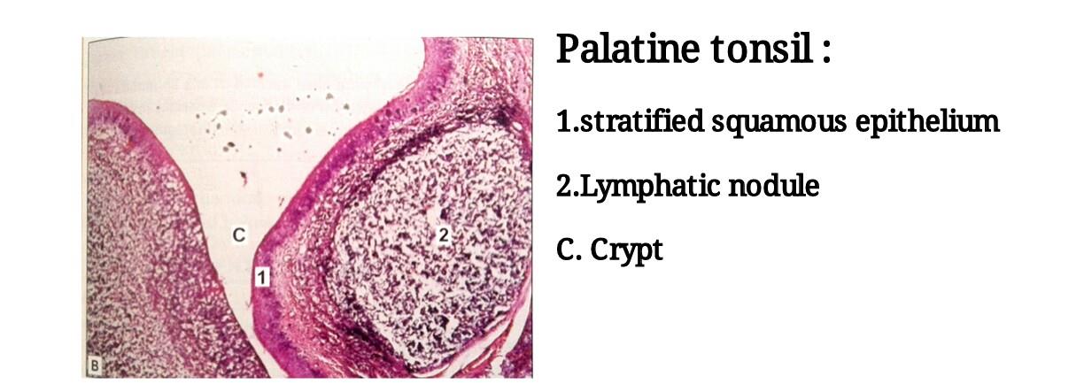
Slide 10: PALATINE TONSIL
DRAWING:
IDEAL SLIDE:
IDENTIFICATION POINTS:
| 1. Palatine tonsil is an aggregation of lymphoid tissue that is readily recognized by the fact that it is covered by a stratified squamous epithelium. |
| 2, At places, the epithelium dips into the tonsil in the form of deep crypts. |
| 3. Deep to the epithelium, there is diffuse lymphoid tissue in which typical lymphatic nodules can be seen. |
Vascular System Histology
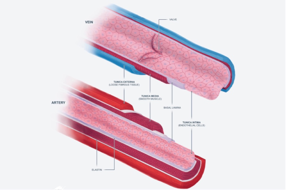
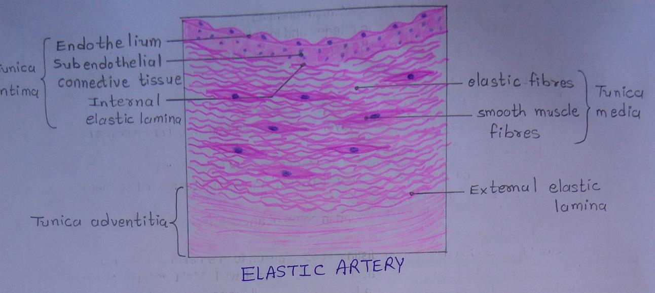
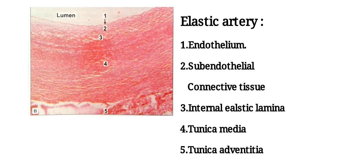
Slide 11: ELASTIC ARTERY
DRAWING:
IDEAL SLIDE:
IDENTIFICATION POINTS:
| 1. Tunica intima consisting of endothelium, subendothelium connective tissue, and internal elastic lamina. |
| 2. The first layer of elastic fibers is called the internal elastic lamina. |
| 3. The internal elastic lamina is not distinct from the elastic fibers of media. |
| 4. Well-developed subendothelial layer in tunica intima. |
| 5. Thick tunica media with many elastic fibers and some smooth muscle fibers. |
| 6. Tunica adventitia containing collagen fibers with several elastic fibers. |
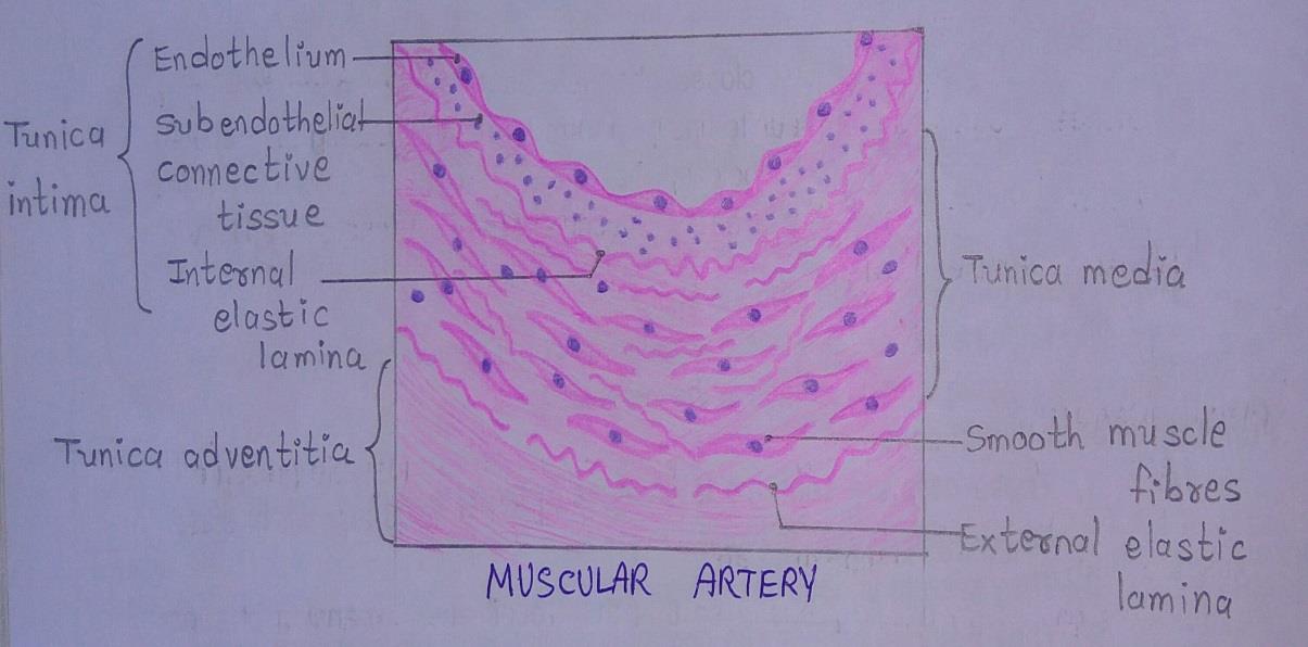
Slide 12: MUSCULAR ARTERY
DRAWING:
IDEAL SLIDE:
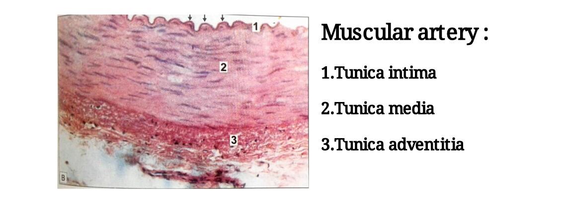
IDENTIFICATION POINTS:
| 1. In the tunica intima, the internal elastic lamina in the muscular arteries stands out distinctly from the muscular media and it is thrown into wavy folds due to contraction of smooth muscle in the media. |
| 2. Tunica media is made up mainly of smooth muscles. |
| 3. Between groups of muscle fibers some connective tissue is present, which may contain some elastic fibers. |
| 4. Tunica adventitia contains collagen fibers and few elastic fibers. |
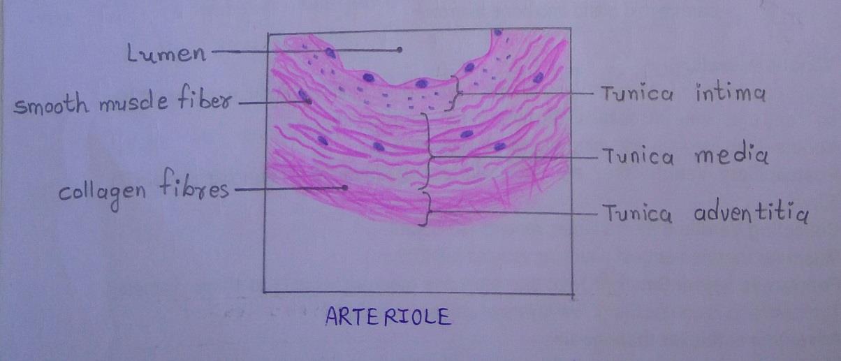
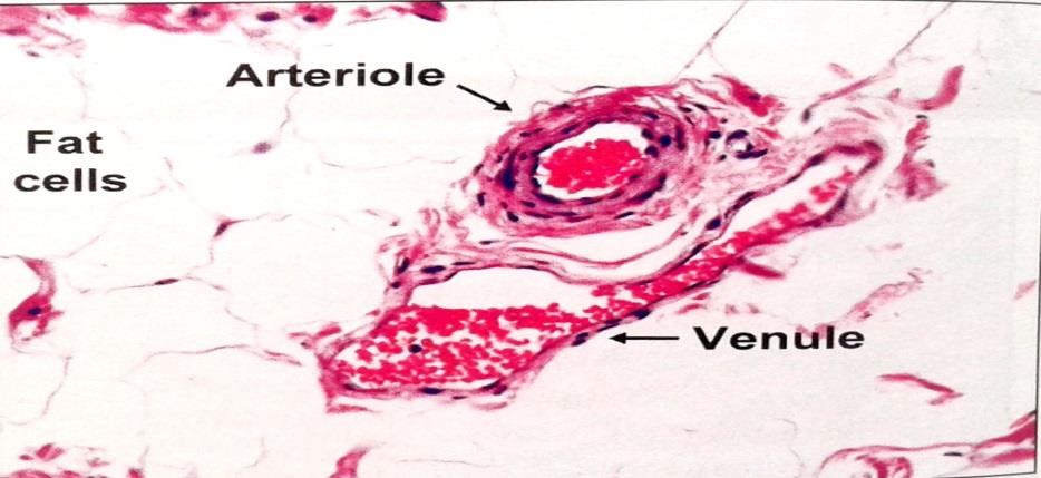
Slide 13: ARTERIOLE
DRAWING:
IDEAL SLIDE:
IDENTIFICATION POINTS:
| 1. Muscular arterioles can be distinguished from true arteries: - By their small diameter. - They do not have an internal elastic lamina. They have a few layers of smooth muscle in their media. |
| 2. Terminal arterioles can be distinguished from muscular arterioles as follow: - They have a diameter of less than 50 micrometer. - They have only a thin layer of muscle in their walls. |
| 3. All the three layers, i.e., tunica adventitia, tunica media, and tunica intima are thin as compared to arteries. |
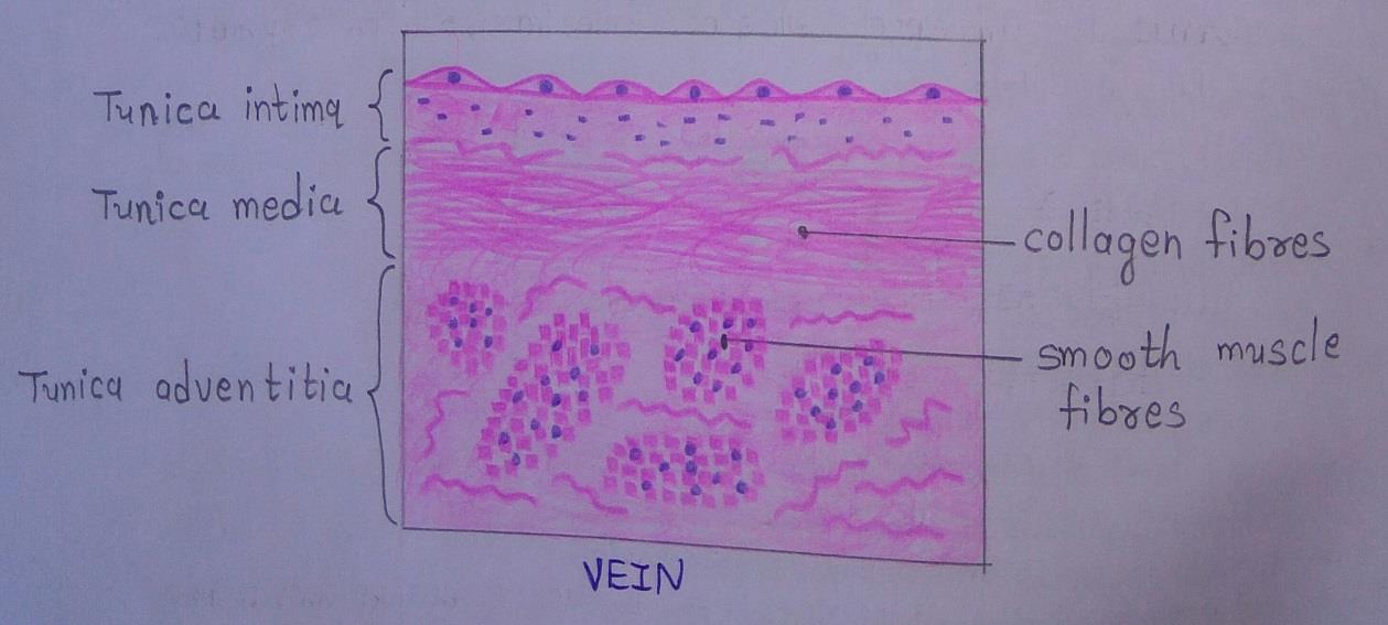
Slide 14: VEIN
DRAWING:
IDEAL SLIDE:
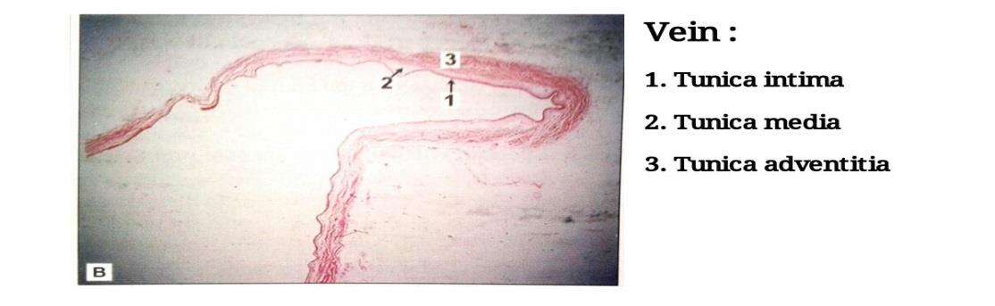
IDENTIFICATION POINTS:
| 1. The tunica media contains a much larger quantity of collagen than in arteries. |
| 2. In arteries, the tunica media is usually thicker than the adventitia. In contrast, the adventitia of veins is thicker than the media. |
| 3. A clear distinction between the tunica intima, media, and adventitia cannot be made out in small veins as all these layers consist predominantly of fibrous tissue. |
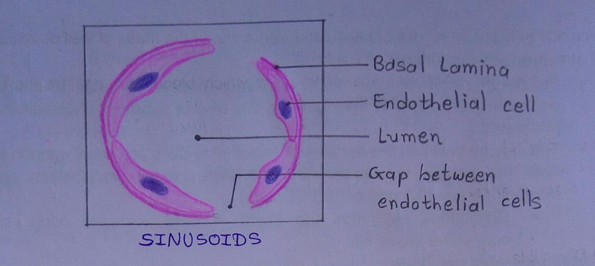
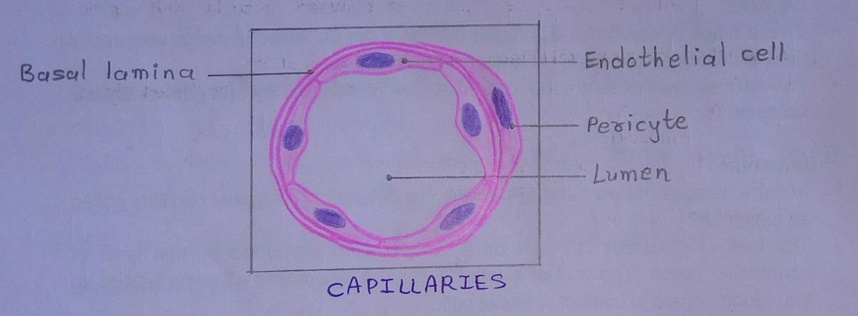
Slide 15: SINUSOIDS & CAPILLARY
DRAWING:
Integumentary System Histology
Slide 16: THIN SKIN
DRAWING:
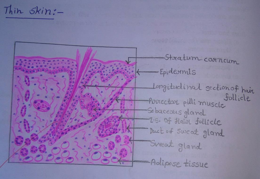
IDEAL SLIDE:
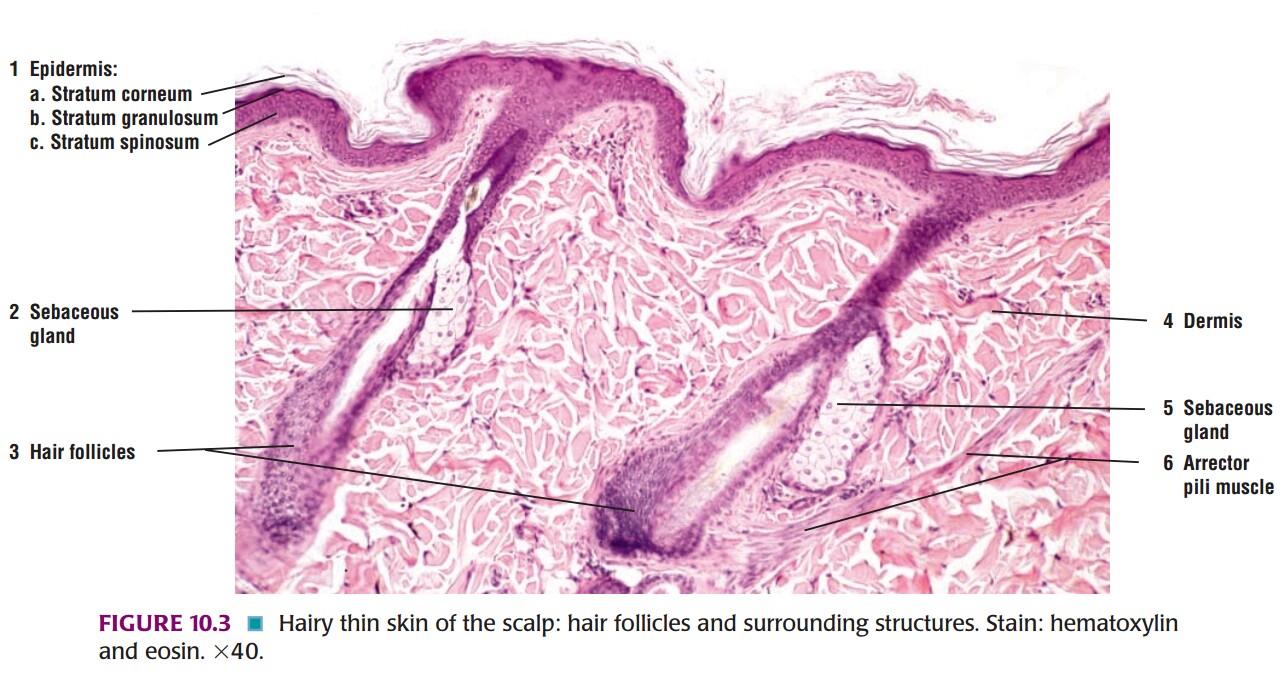
IDENTIFICATION POINTS:
| 1. Stratum corneum of epidermis is thin. |
| 2. Stratum lucidum of epidermis is absent. |
| 3. Epidermal ridges are absent. |
| 4. Hair follicles, arrector pilli muscle, and sebaceous glands are present. |
| 5. Sweat glands in the dermis are few. |
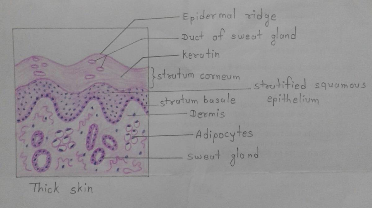
Slide 17: THICK SKIN
DRAWING:
IDEAL SLIDE:
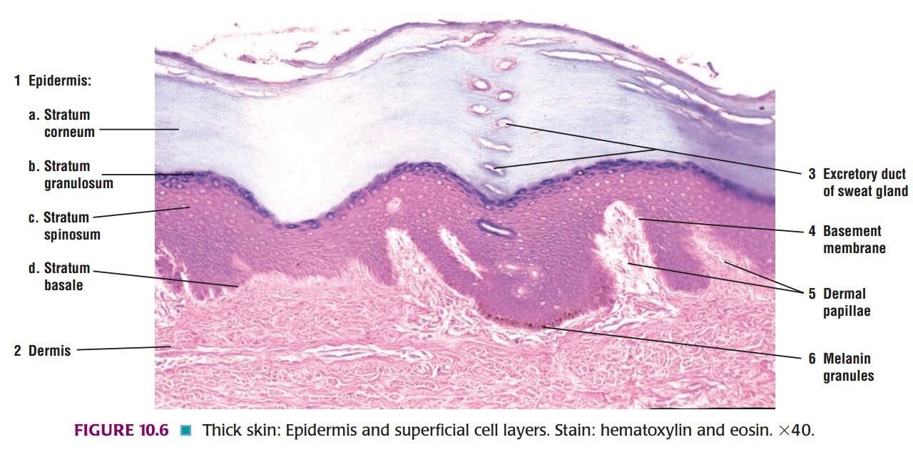
IDENTIFICATION POINTS:
| 1. Stratum corneum of the epidermis is very thick. |
| 2. Stratum lucidum of epidermis is present. |
| 3. Epidermal ridges are well developed. |
| 4. Hair follicles, arrector pilli muscle, and sebaceous glands are absent. |
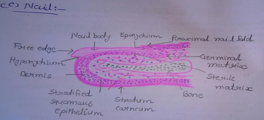
Slide 18: NAIL
DRAWING:
Comments (0)