Respiratory & Glands (Systemic Histology 1)
6 years ago 31050
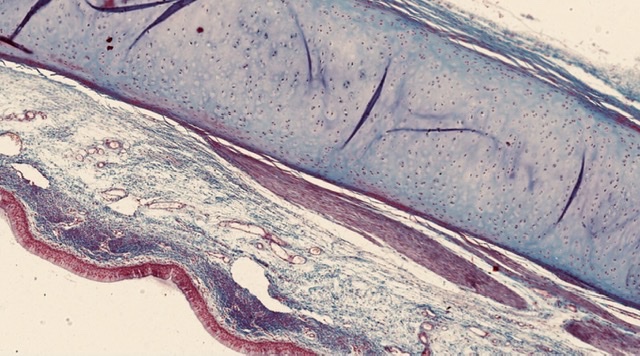
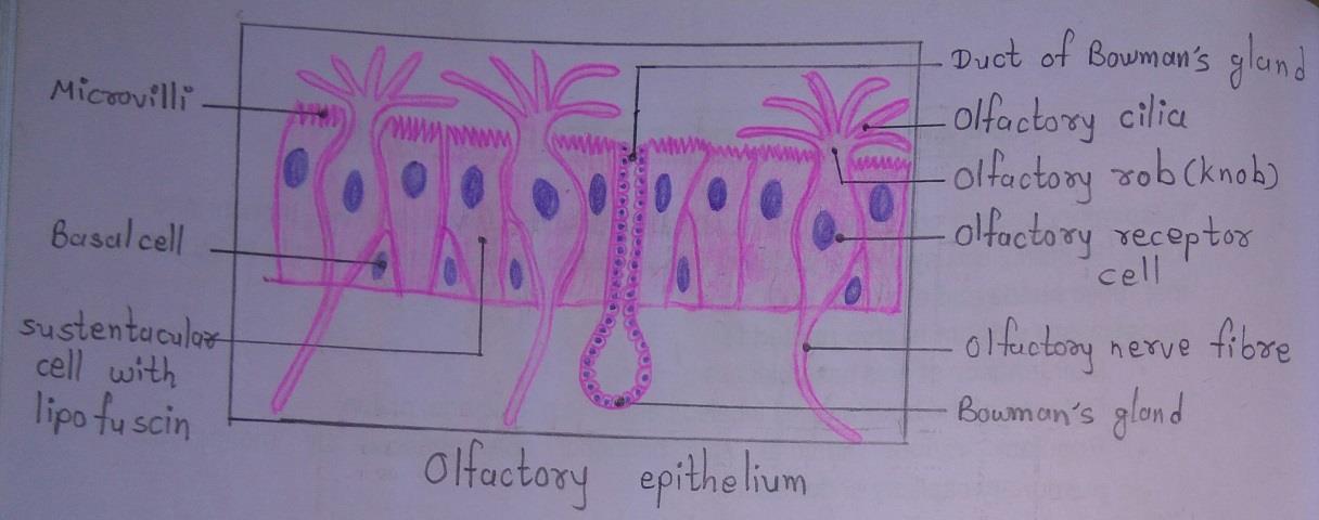
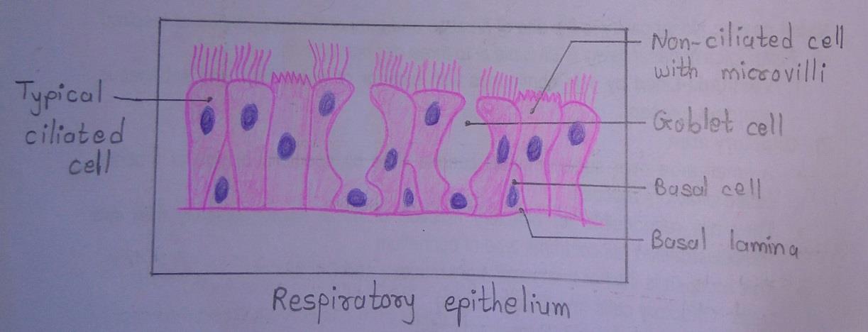
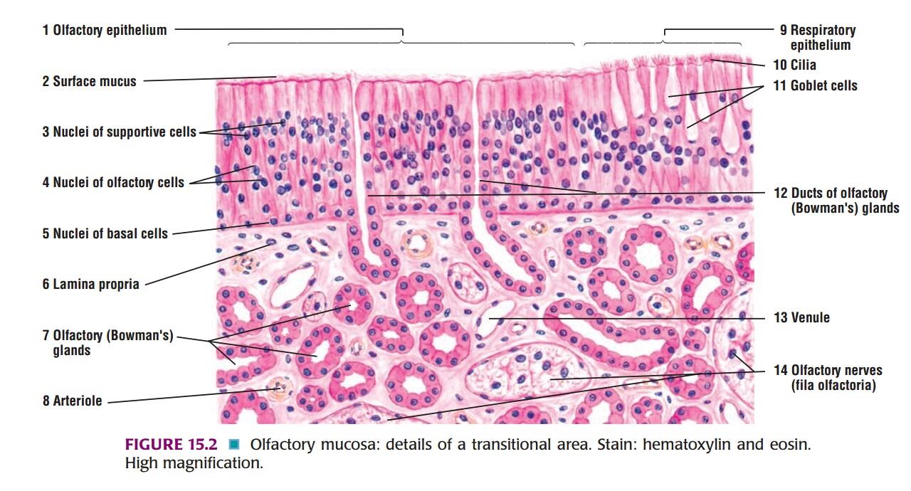
Slide 1: OLFACTORY & RESPIRATORY EPITHELIUM
DRAWING:
IDEAL SLIDE:
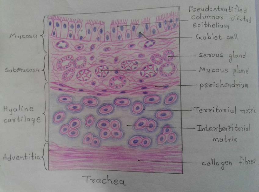
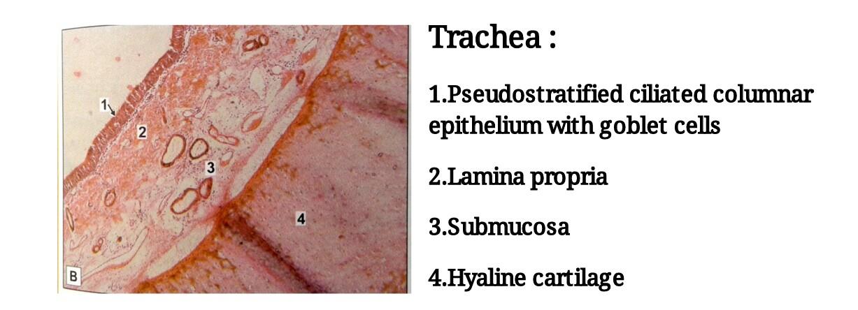
Slide 2: TRACHEA
DRAWING:
IDEAL SLIDE:
IDENTIFICATION POINTS:
| 1. Mucosa is formed by pseudostratified ciliated columnar epithelium with goblet cells, basal cells and the underlying lamina propria. |
| 2. Submucosa made up of loose connective tissue containing mucous gland, serous gland and numerous aggregations of lymphoid tissue. |
| 3. C-shaped mass of hyaline cartilage is present. |
| 4. The connective tissue in the wall of the trachea contains many elastic fibers. |
| 5. Adventitia is made of fibroelastic connective tissue containing blood vessels and nerves. |
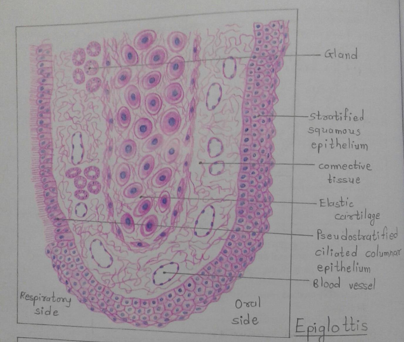
Slide 3: EPIGLOTTIS
DRAWING:
IDENTIFICATION POINTS:
| 1. The surface of epiglottis is covered on the oral side by stratified squamous epithelium and on the respiratory side by pseudostratified ciliated columnar epithelium. |
| 2. The core of the epiglottis is made up of a plate of elastic cartilage covered by connective tissue in which there are numerous blood vessels and mucous glands. |
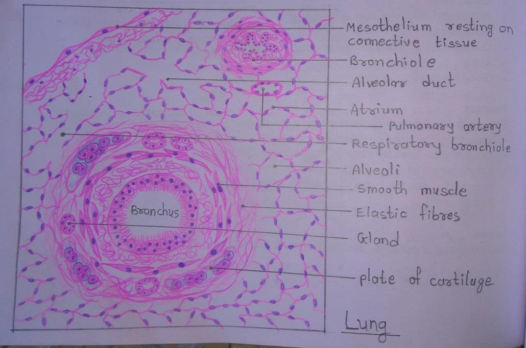
Slide 4: LUNG
DRAWING:
IDEAL SLIDE:
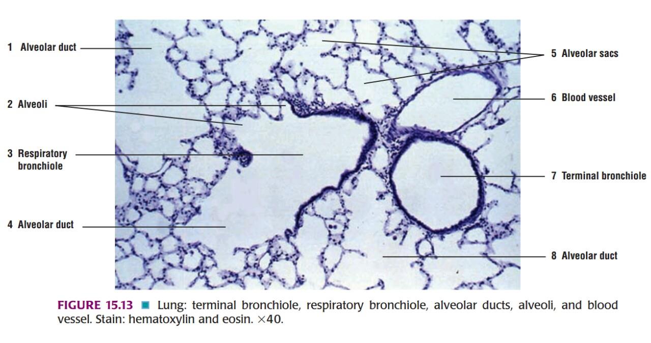
IDENTIFICATION POINTS:
| 1. The lung surface is covered by pleura. It consists of a lining of mesothelium resting on a layer of connective tissue. |
| 2. The lung parenchyma is made up of numerous thin-walled spaces or alveoli. |
| 3. The alveoli give a honeycomb appearance and are lined by flattened squamous cells. They are filled with air. |
| 4. The intrapulmonary bronchus is lined by pseudostratified ciliated columnar epithelium with few goblet cells. Its structure is similar to the trachea i.e. it has smooth muscles, cartilage, and glands present in its wall. |
| 5. The bronchiole is lined by simple columnar or cuboidal epithelium surrounded by bundles of smooth muscle cells. |
| 6. Bronchioles subdivide and when their diameter is approximately 1mm or less, they are called terminal bronchiole. |
| 7. Respiratory bronchiole, alveolar duct and atrium are also present. |
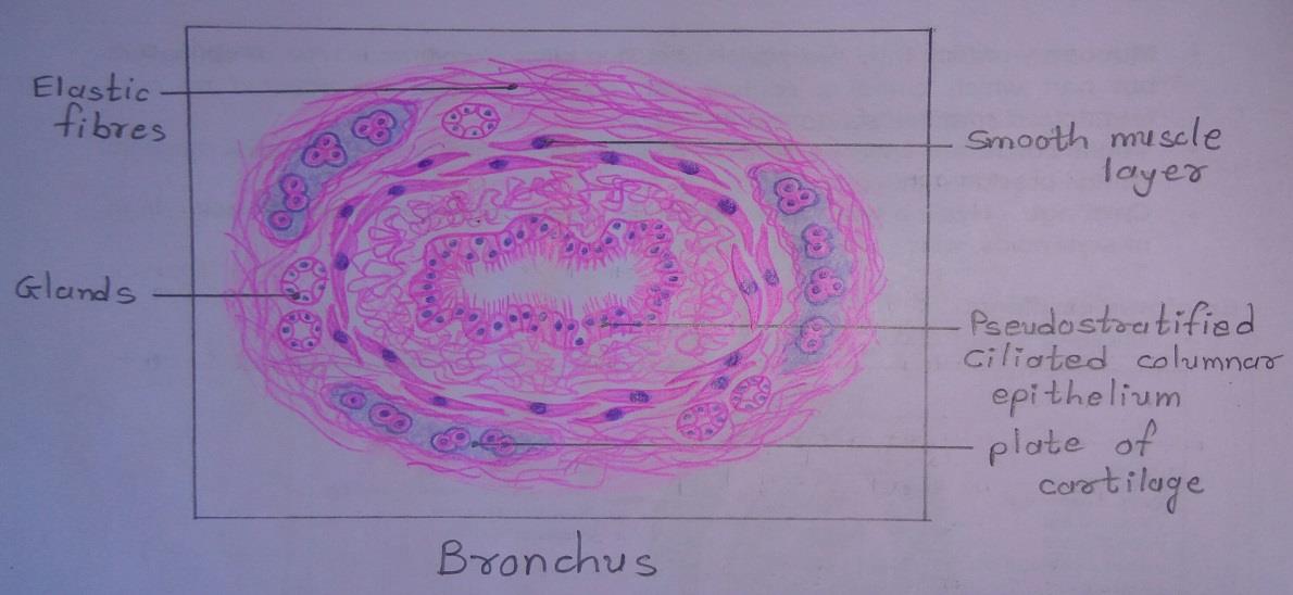
Slide 5: BRONCHUS
DRAWING:
IDEAL SLIDE:
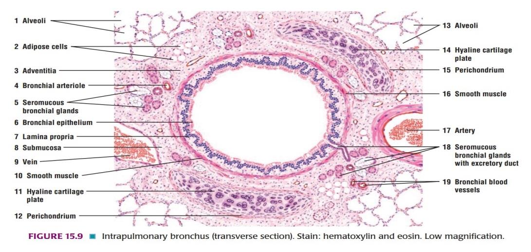
IDENTIFICATION POINTS:
| 1. Bronchus is lined by pseudostratified ciliated columnar epithelium. |
| 2. The cartilage in the wall of the bronchus becomes irregular in shape and is progressively smaller. |
| 3. The amount of muscle in the bronchial wall increases as the bronchus becomes smaller. |
| 4. Both serous and mucous acini present between the cartilage and muscle layer. |
Glands Histology
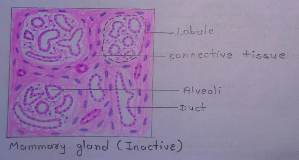
Slide 6: INACTIVE MAMMARY GLAND
DRAWING:
IDEAL SLIDE:
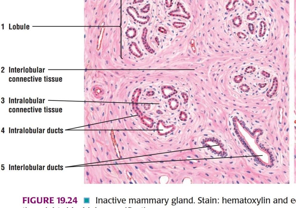
IDENTIFICATION POINTS:
| 1. Mammary gland consists of lobules of glandular tissue separated by a considerable quantity of connective tissue and fat. |
| 2. Non-lactating mammary glands contain more connective tissue and less glandular tissue. |
| 3. The glandular elements or alveoli are distinctly tubular. They are lined by cuboidal epithelium and have a large lumen so that they look like ducts. Some of them may be in form of solid cords of cells. |
| 4. Extensive branching of duct system seen. |
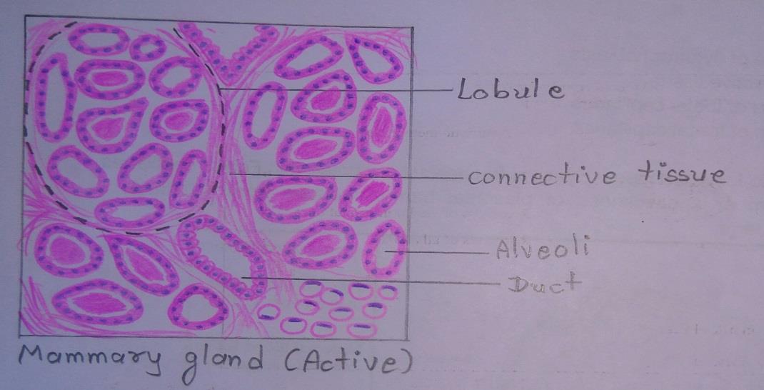
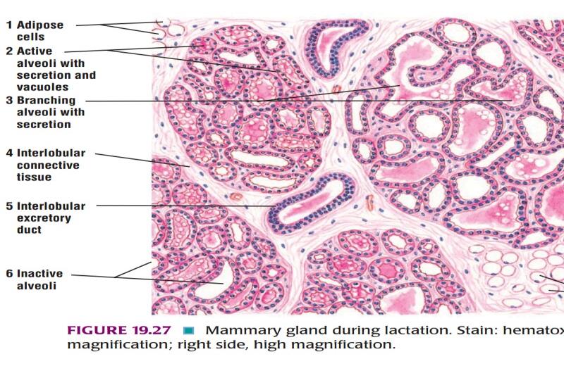
Slide 7: ACTIVE MAMMARY GLAND
DRAWING:
IDEAL SLIDE:
IDENTIFICATION POINTS:
| 1.In the lactating mammary gland, the glandular elements proliferate so that they become relatively more prominent than the connective tissue. |
| 2.The interlobular connective tissue septum is very thin. |
| 3.The lobules are formed by compactly arranged alveoli. |
| 4.The alveoli are lined by simple cuboidal secretory epithelium and associated myoepithelial cells. Their lumen contains eosinophilic secretory material which appear vacuolated due to the presence of fat droplets. |
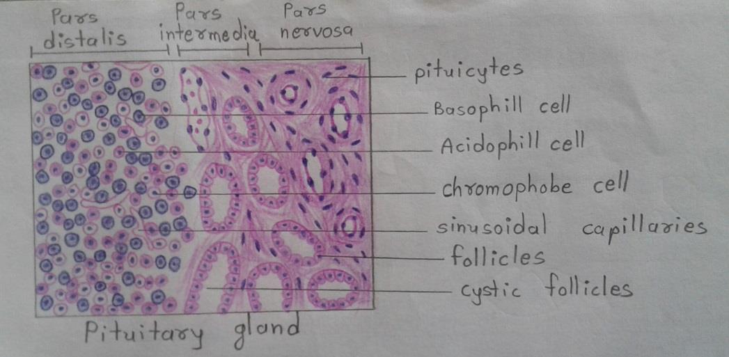
Slide 8: PITUITARY GLAND
DRAWING:
IDEAL SLIDE:
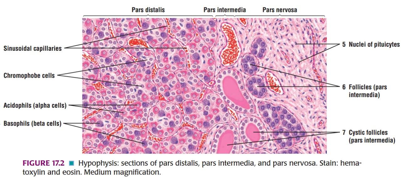
IDENTIFICATION POINTS:
| 1. The pars anterior of the hypophysis cerebri consists of cells separated by fenestrated sinusoids. |
| 2. The cells are of three types • The pink staining cells are alpha cells or acidophils. • The cells with bluish cytoplasm are beta cells or basophils. • Cells in which the cytoplasm is not conspicuous, and the nuclei are closely packed, are chromophobe cells. |
| 3. The pars intermedia is poorly developed in the human hypophysis. |
| 4. In ordinary preparations the most conspicuous feature is the presence of colloid filled vesicles. These vesicles are remnants of the pouch of Rathke. |
| 5. The pars posterior consists of numerous unmyelinated nerve fibers which are the axons of neurons located in the hypothalamus. |
| 6. Situated between these axons there are supporting cells of a special type called pituitocytes. |
| 7. The collection of secretory granules at the terminal portion of axonal processing is called as Herring bodies. |
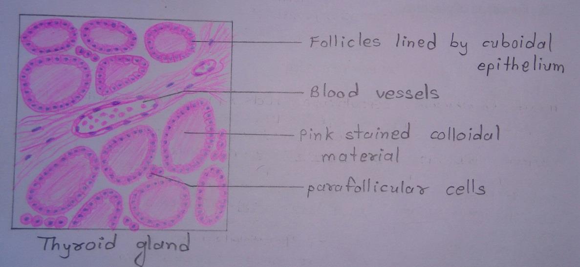
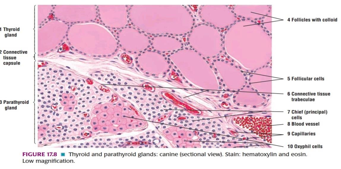
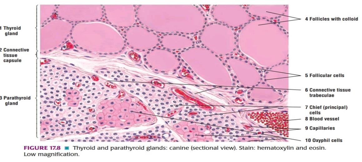
Slide 9: THYROID GLAND
DRAWING:
IDEAL SLIDE:
IDENTIFICATION POINTS:
| 1. The thyroid gland is made up of follicles lined by cuboidal epithelium. |
| 2. In photomicrographs in low magnification it can be seen that follicles vary in shape and size. |
| 3. Each follicle is filled with a homogenous pink colloid proteinaceous material composed primarily of thyroglobulin that has been produced by the follicular epithelial cells. |
| 4. Parafollicular cells are present in relation to the follicles and also as groups in the connective tissue. |
| 5. Note the blood vessels between follicles. |
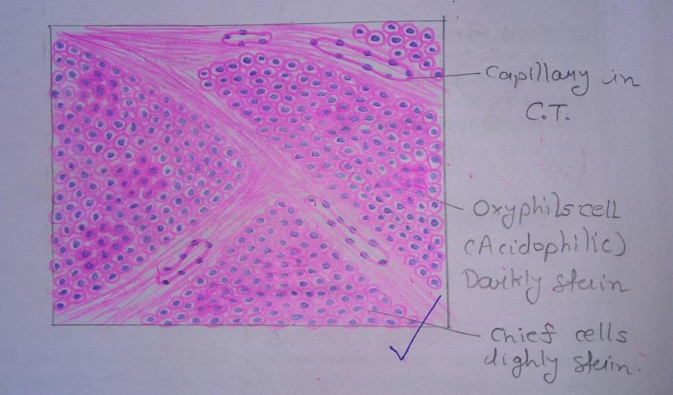
Slide 10: PARATHYROID GLAND
DRAWING:
IDEAL SLIDE:
IDENTIFICATION POINTS:
| 1. The cells of parathyroid glands are of two main types • Chief cells or principal cells. • Oxyphil cells or eosinophil cells. |
| 2. The chief cells are much more numerous than the oxyphil cells. |
| 3. The chief cells are seen to be small round cells with vesicular nuclei. Their cytoplasm is clear and either mildly eosinophil or basophil. |
| 4. The oxyphil cells are much larger than the chief cells and contain granules that stain strongly with acid dyes. Their nuclei are smaller and stain more intensely than those of chief cells. |
| 5. Adipose cells are also seen. |
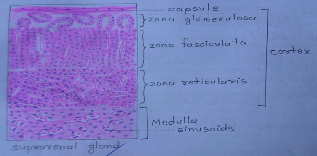
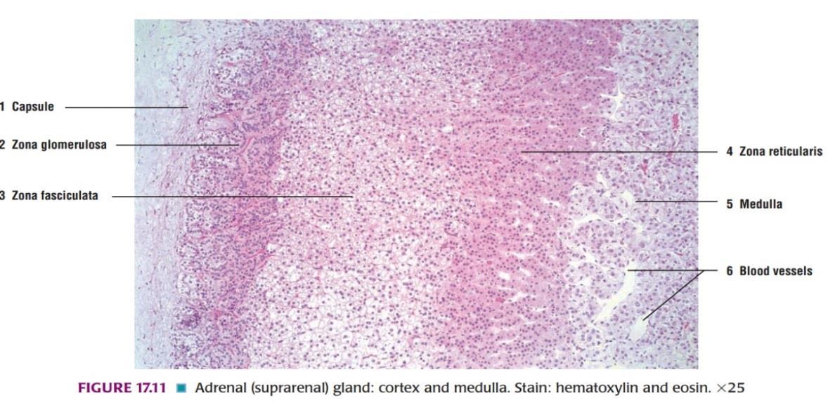
Slide 11: SUPRARENAL GLAND
DRAWING:
IDEAL SLIDE:
IDENTIFICATION POINTS:
| 1) In zona glomerulosa, the cells are arranged as inverted U-shaped formations. The cells of the zona glomerulosa are seen to be small, polyhedral, or columnar, with basophilic cytoplasm and deeply staining nuclei. |
| 2) In zona fasciculata, the cells are arranged in straight columns, two cells thick. Sinusoids intervene between the columns. The cells of the zona fasciculata are seen to be large, polyhedral, with basophilic cytoplasm and vesicular nuclei. |
| 3) The cells in zona reticularis are smaller and more acidophilic than the other two-layer. The zona reticularis is made up of cords of cells that branch and form a network. |
| 4) The medulla of the suprarenal gland is made up of chromaffin cells. The chromaffin cells are arranged in groups or columns which are separated by wide sinusoids. |
Comments (0)