Digestive Viscera (Systemic Histology 2)
6 years ago 22532
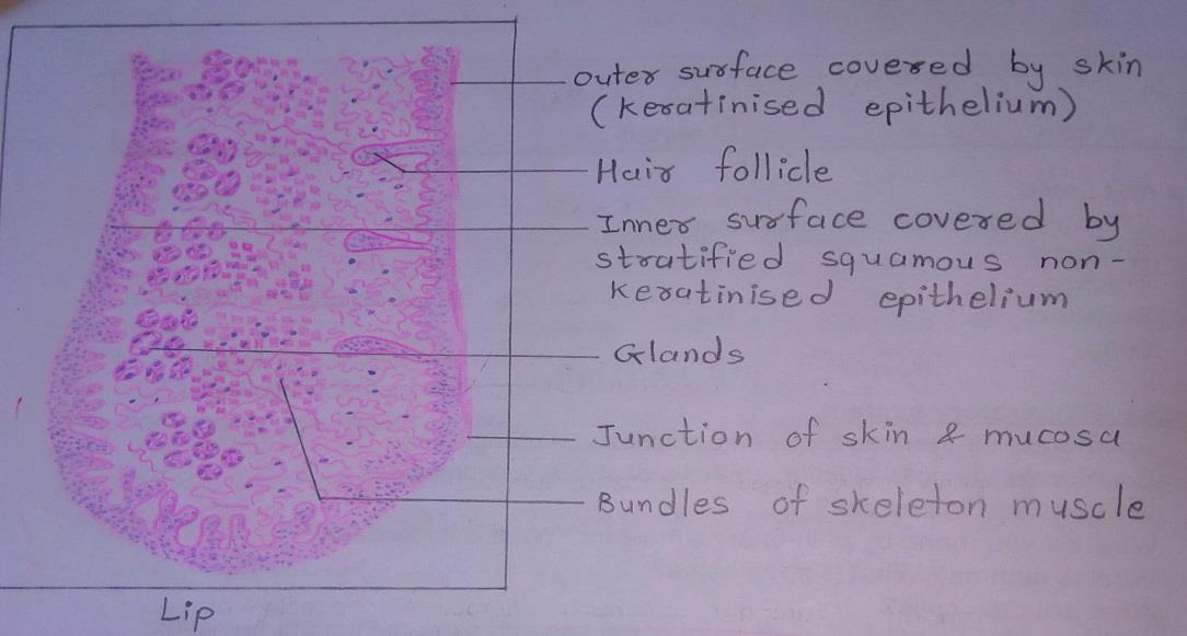
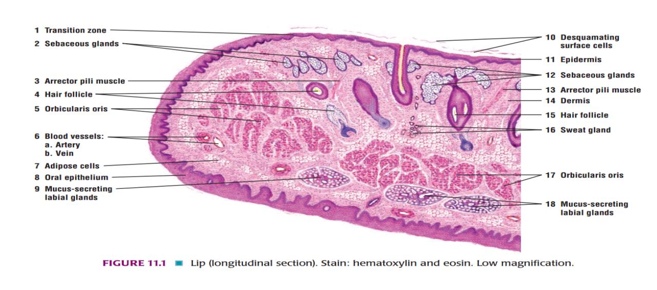
Slide 1: LIP
DRAWING:
IDEAL SLIDE:
IDENTIFICATION POINTS:
| 1. The substance of the lip is formed by a mass of muscle. |
| 2. Each lip has an ‘external’ surface covered by skin and an ‘internal’ surface lined by mucous membrane. |
| 3. The ‘external’ surface of the lip is lined by true skin in which hair follicles and sebaceous glands can be seen. |
| 4. The mucous membrane is lined by stratified squamous non-keratinized epithelium. |
| 5. The epithelium has a well-marked rete ridge system. The term rete ridges are applied to finger-like projections of epithelium that extend into underlying connective tissue, just like the epidermal papillae. |
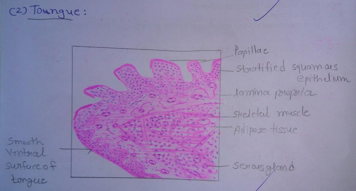
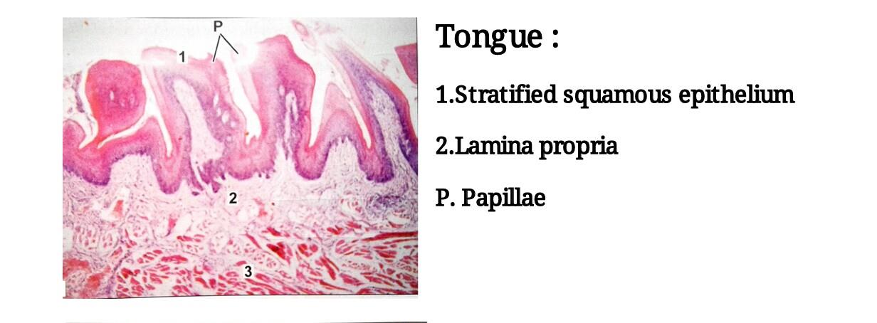
Slide 2: TONGUE
DRAWING:
IDEAL SLIDE:
IDENTIFICATION POINTS:
| 1. The tongue is covered on both surfaces by non-keratinized stratified squamous epithelium. |
| 2. The ventral surface of the tongue is smooth, but on the dorsum, the surface shows numerous projections or papillae. |
| 3. Each papilla has a core of connective tissue covered by epithelium. Some papillae are pointed(filiform), while others are broad at the top (fungiform). A third type of papilla is circumvallate, the top of this papilla is broad and lies at the same level as the surrounding mucosa. |
| 4. The main mass of the tongue is formed by skeletal muscle seen below the lamina propria. Muscle fibers run in various directions so that some are cut longitudinally and some transversely. |
| 5. Numerous serous and mucous glands are present amongst the muscle fibers. |
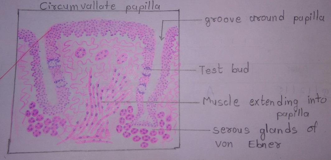
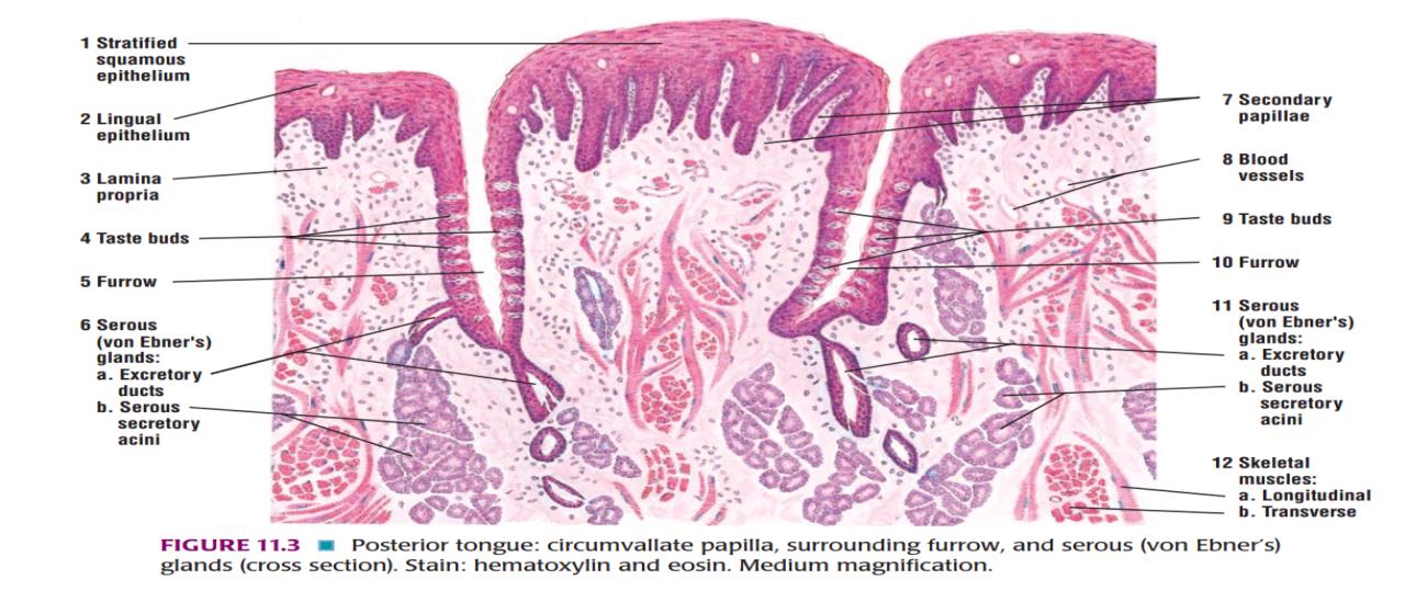
Slide 3: CIRCUMVALLATE PAPILLA
DRAWING:
IDEAL SLIDE:
IDENTIFICATION POINTS:
| 1. In sections through the papilla it is seen that the papilla has a circumferential ‘lateral wall’ that lies in the depth of the groove. |
| 2. They are characterized by their dome-shaped structure lined stratified squamous epithelium. |
| 3. Numerous ovals shaped lightly stained taste buds can be seen on the lateral wall of the papillae. |
| 4. Ducts of serous gland of Von Ebner is open in groove around the papilla. |
| 5. Skeleton muscle can be seen extending into the papillae. |
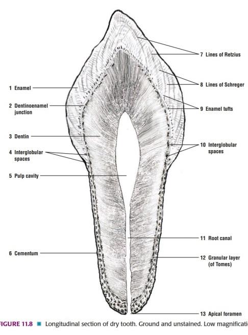
Slide 4: GROUND SECTION OF TEETH
IDEAL SLIDE:
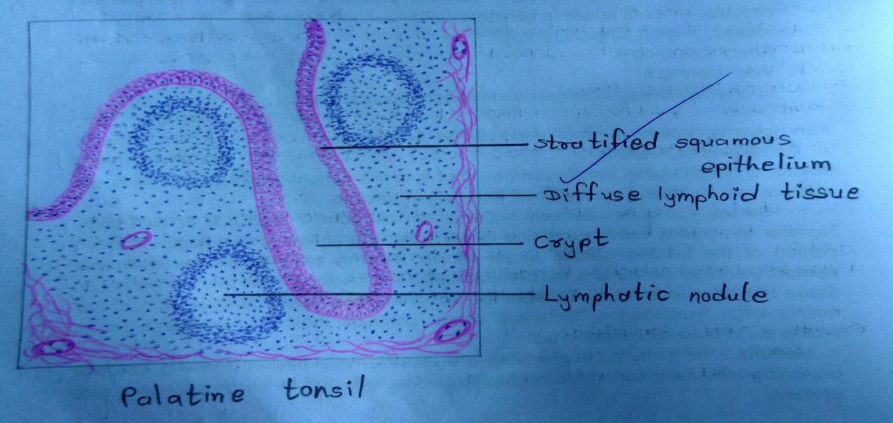
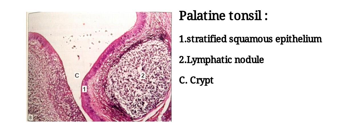
Slide 5: PALATINE TONSIL
DRAWING:
IDEAL SLIDE:
IDENTIFICATION POINTS:
| 1. Palatine tonsil is an aggregation of lymphoid tissue that is readily recognized by the fact that it is covered by a stratified squamous epithelium. |
| 2. At places the epithelium dips into the tonsil in the form of deep crypts. |
| 3. Deep to the epithelium, there is diffuse lymphoid tissue in which typical lymphatic nodules can be seen. |
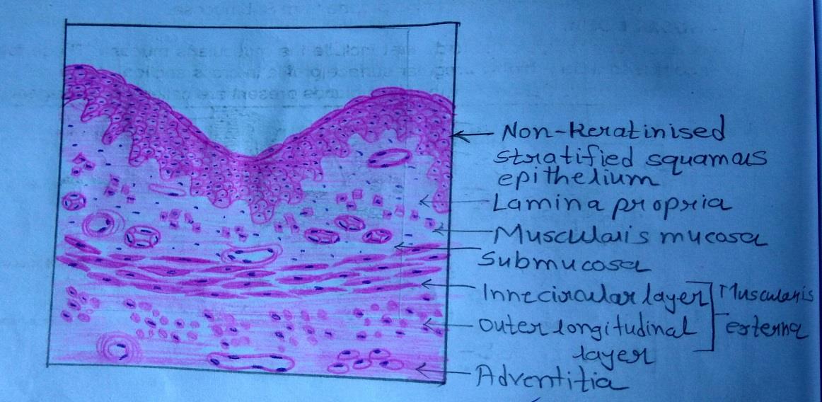
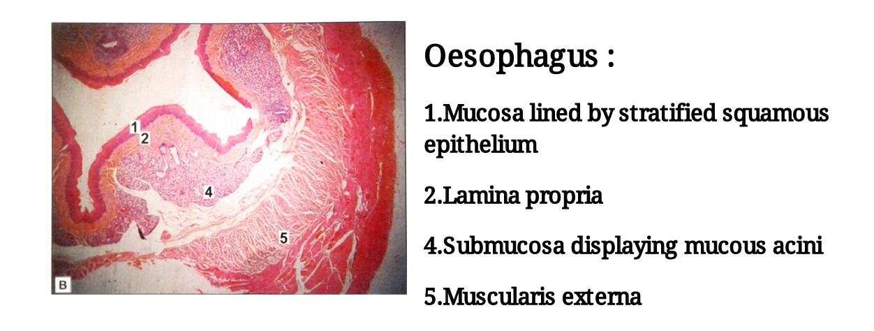
Slide 6: OESOPHAGUS
DRAWING:
IDEAL SLIDE:
IDENTIFICATION POINTS:
| 1. The mucous membrane of the esophagus shows several longitudinal folds. |
| 2. The mucosa is lined by non-keratinized stratified squamous epithelium. |
| 3. Finger-like processes of the connective tissue of the lamina propria project into the epithelial layer. |
| 4. At the upper and lower ends of the esophagus some tubuloalveolar mucous glands are present in the lamina propria. |
| 5. The muscularis mucosae is absent or poorly developed in the upper part of the esophagus. It is distinct in the lower part of the esophagus. |
| 6. The only special feature of the submucosa is the presence of compound tubuloalveolar mucous glands. Small aggregations of lymphoid tissue may be present in the submucosa. |
| 7. The muscle layer consists of the usual circular and longitudinal layers. However, it is unusual in that the muscle fibers are partly striated and partly smooth. |
| 8. The muscle layer of the esophagus is surrounded by dense fibrous tissue that forms an adventitial coat for the esophagus. |
Slide 7: CARDIAC PART OF STOMACH
DRAWING:
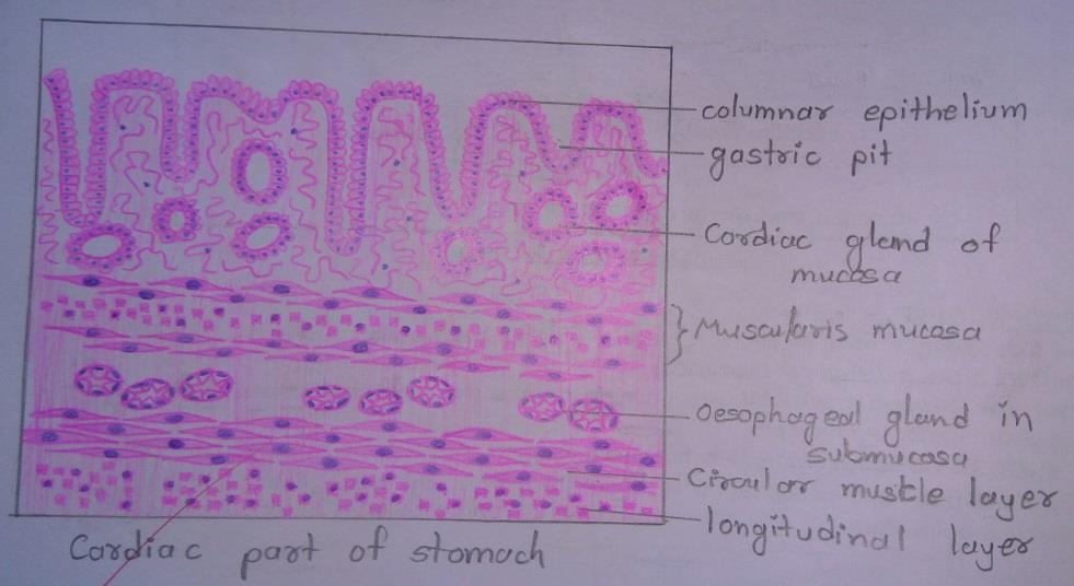
IDENTIFICATION POINTS:
Slide 8: FUNDUS PART OF STOMACH
DRAWING:
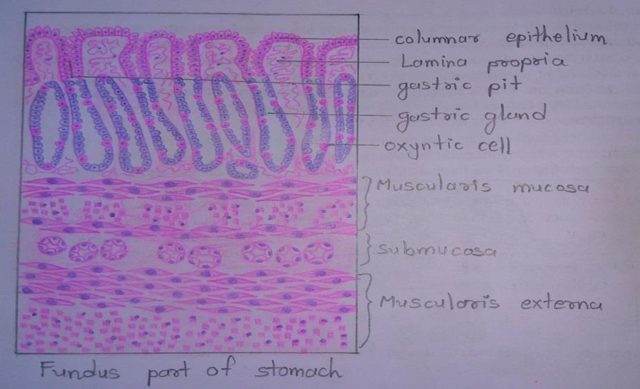
IDEAL SLIDE:
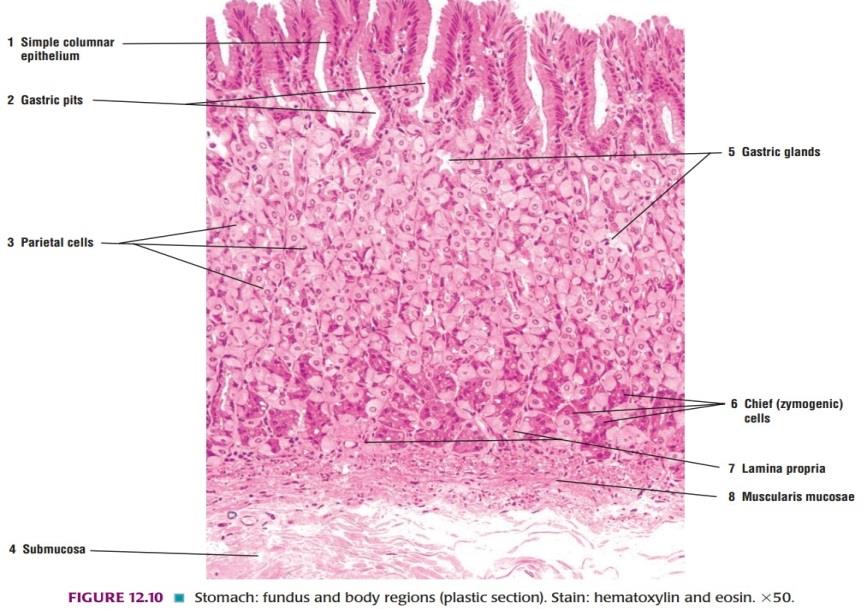
IDENTIFICATION POINTS:
| 1. Mucosa is lined by simple tall columnar epithelium. It shows invaginations called gastric pits that occupy the superficial one-fourth of the mucosa. |
| 2. The area between pits and the muscularis mucosae is packed with tubular gastric glands. |
| 3. The glands are lined mainly by blue staining chief cells or peptic cells. Amongst these, there are pink staining oxyntic cells. These are large cells that are placed peripherally in the wall of the gland. They are more numerous in the upper parts of the gastric glands. |
| 4. Muscularis externa is composed of three layers of smooth muscle- inner oblique, middle circular and outer longitudinal. |
| 5. Observe that the gastric pits occupy the upper one-fourth of the lamina propria of the mucosa. |
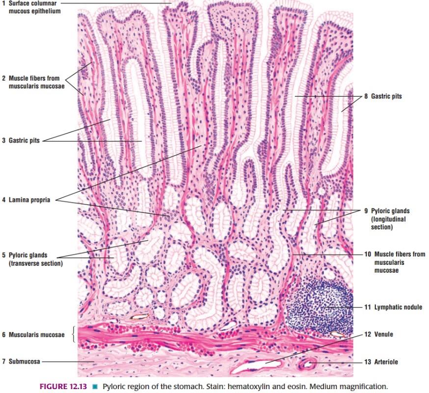
Slide 9: PYLORIC PART OF THE STOMACH
DRAWING:
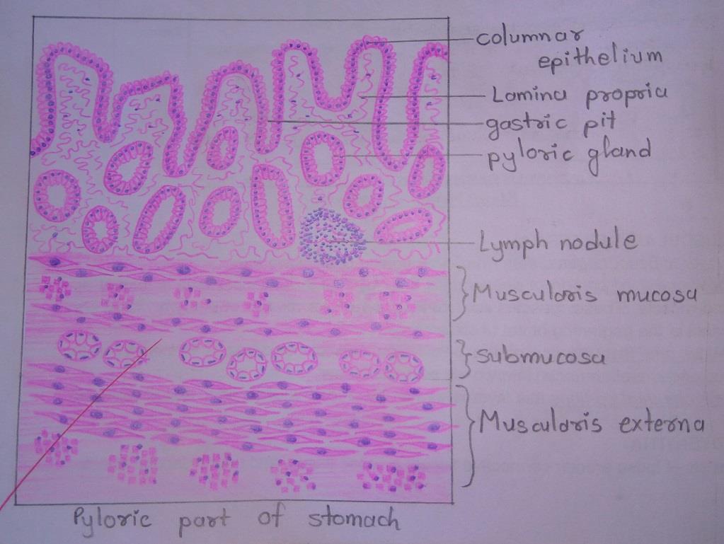
IDEAL SLIDE:
IDENTIFICATION POINTS:
| 1. In the pyloric part of the stomach the gastric pits are much deeper than in the body of the stomach. |
| 2. Deep to the pits, there are pyloric glands that are lined by mucous secreting cells. These are pale staining. |
| 3. The muscularis mucosae, submucosa, and part of the muscle coat are also seen. |
| 4. It is important to note that the stomach does not have villi. In the photomicrograph folds of epithelial lining may be confused with villi. |
| 5. Observe that each fold merges with underlying connective tissue completely. When true villi are present, small parts of them appear as circular or oval masses not attached to a villus or to the submucosa. these are villi that have been cut transversely or obliquely. |
| 6. Another important feature to note is that the lining epithelium does not have typical goblet cells, but some epithelial cells are mucous secreting. |
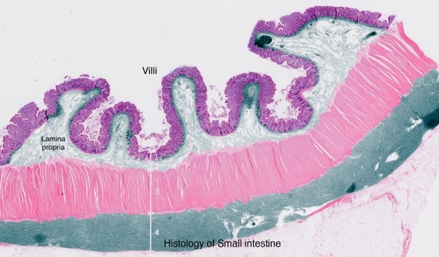
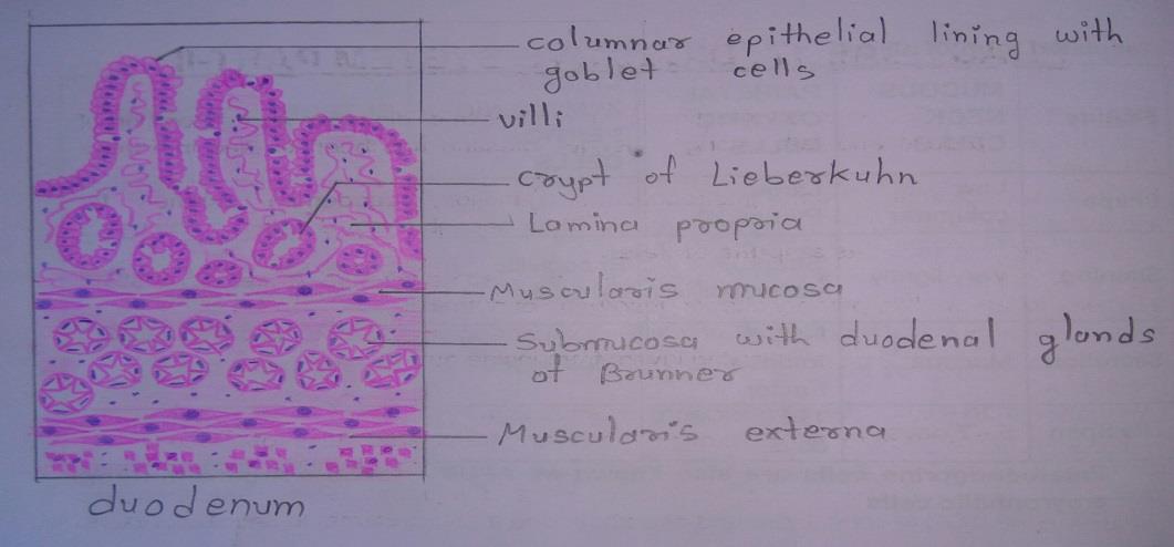
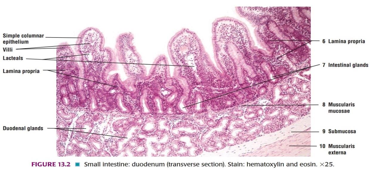
Slide 10: DUODENUM
DRAWING:
IDEAL SLIDE:
IDENTIFICATION POINTS:
| 1. The mucosa consists of: • Numerous finger-like processes, or villi, that project from the surface of the mucosa into the lumen. • Numerous depressions or crypts that invade the lamina propria. |
| 2. The duodenum is easily distinguished from the jejunum or ileum because of the presence of glands in the submucosa. (No glands are present in the submucosa of the jejunum or ileum) |
| 3. These duodenal glands of Brunner are compound tubule-alveolar glands. |
| 4. Their ducts pass through the muscularis mucosae to open into the intestinal crypts of Lieberkühn. |
| 5. The cells lining the alveoli of duodenal glands are predominantly mucous secreting columnar cells having flattened basal nuclei. |
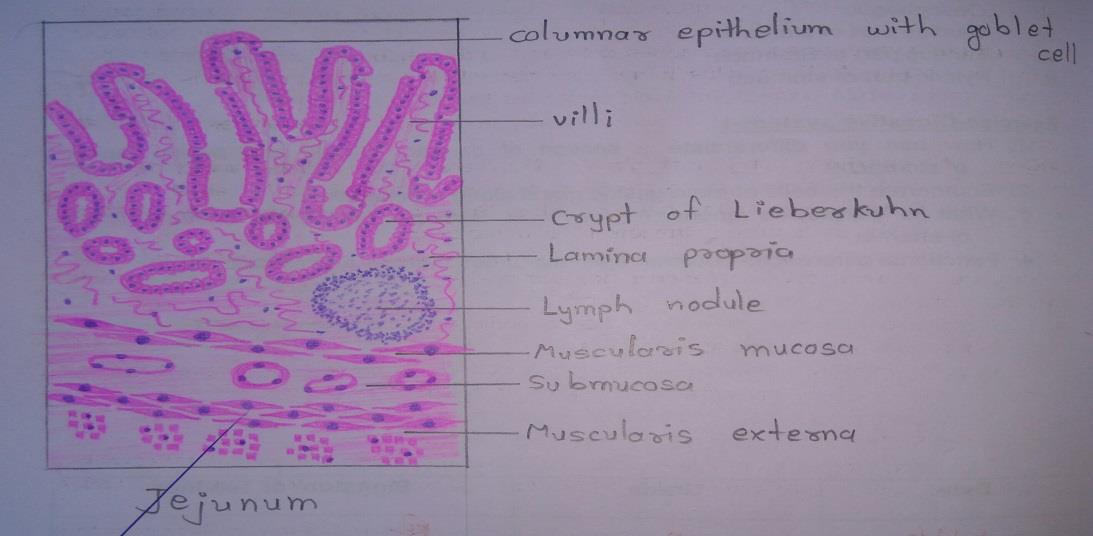
Slide 11: JEJUNUM
DRAWING:
IDEAL SLIDE:
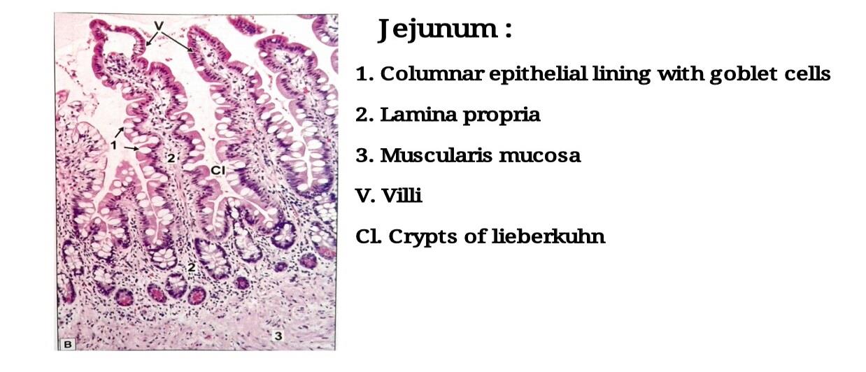
IDENTIFICATION POINTS:
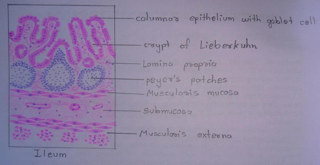
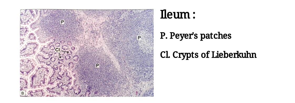
Slide 12: ILEUM
DRAWING:
IDEAL SLIDE:
IDENTIFICATION POINTS:
| 1. The general structure of the ileum is similar to that of the jejunum except for: ≡ The entire thickness of the lamina propria is infiltrated with lymphocytes amongst which typical lymphatic follicles can be seen which may extend into the submucosa. These lymphatic follicles are called as Peyer`s patches. ≡ In the region overlying the Peyer`s patch villi may be rudimentary or absent. |
| 2. The villi are thin and slender in the region of ileum. |
| 3. M cells are found overlying the lymphoid follicles. |
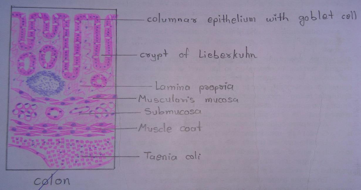
Slide 13: COLON
DRAWING:
IDEAL SLIDE:
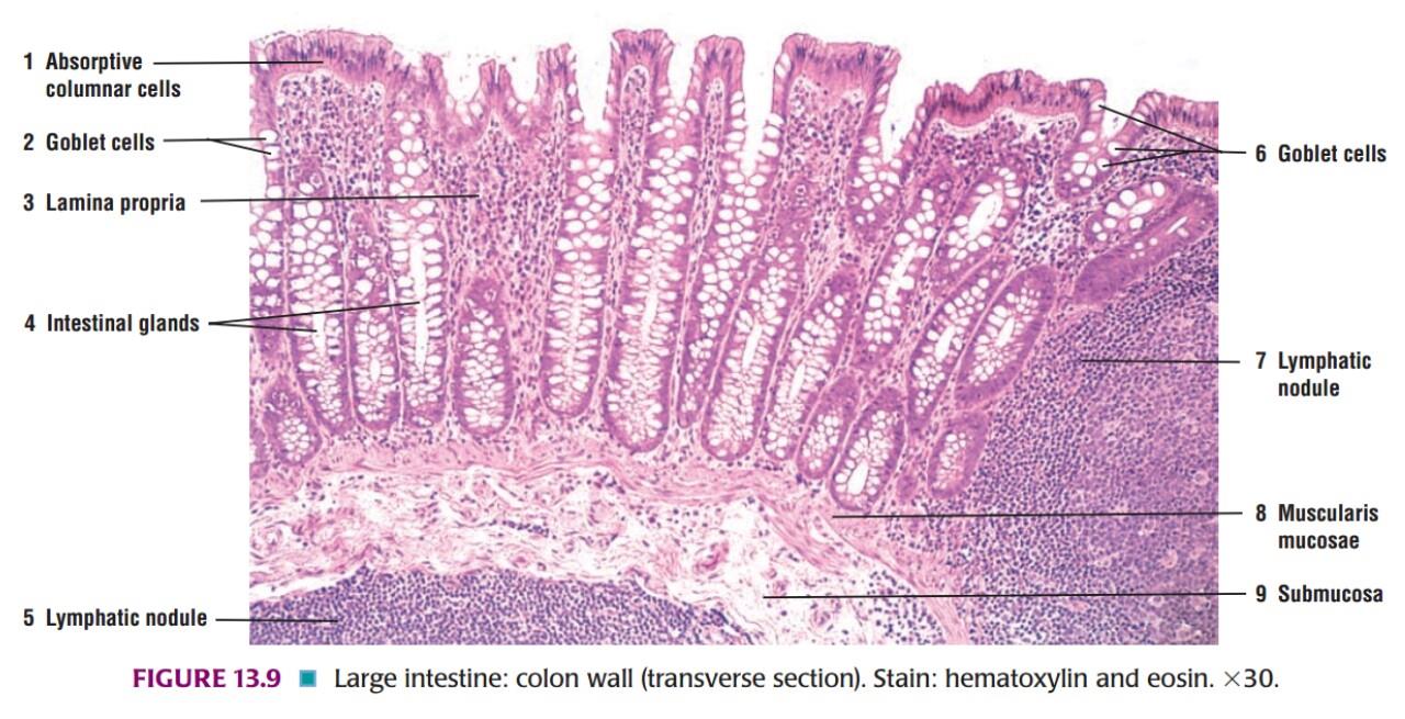
IDENTIFICATION POINTS:
| 1. The most important feature to note is the absence of villi. |
| 2. The mucosa shows numerous tubular glands or crypts. The surface of the mucosa, and the crypts, are lined by columnar cells amongst which there are numerous goblet cells. |
| 3. A section of the large intestine is easily distinguished from that of the small intestine because of the absence of villi; and from the stomach because of the presence of goblet cells (which are absent in the stomach) |
| 4. The muscularis mucosa, submucosa, and circular muscle coat are similar to those in the small intestine. |
| 5. However, the longitudinal muscle coat is gathered into three thick bands called taenia coli. The longitudinal muscle is thin in the intervals between the taenia. |
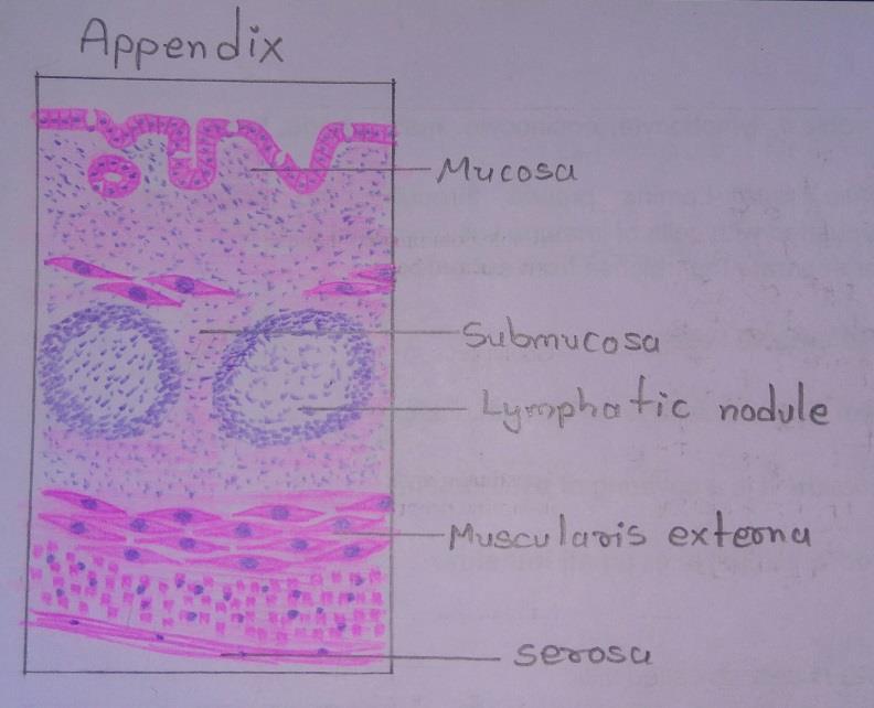
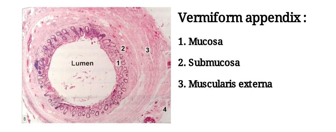
Slide 14: APPENDIX
DRAWING:
IDEAL SLIDE:
IDENTIFICATION POINTS:
| 1. The appendix is the narrowest part of the gastrointestinal canal and is seen as a tubular structure. |
| 2. The innermost layer of the mucosa, is lined by simple columnar epithelium with goblet cells. |
| 3. The crypts are poorly formed. |
| 4. Scattered lymphocytes and aggregated nodules are present in the lamina propria and they may extend into the next layer. |
| 5. The next layer, submucosa may show a variable number of lymphatic nodules. |
| 6. The submucosa is surrounded by a smooth muscle layer followed by serosa. |
| 7. The longitudinal muscle coat is complete and equally thick all around. Taenia coli is not present. |
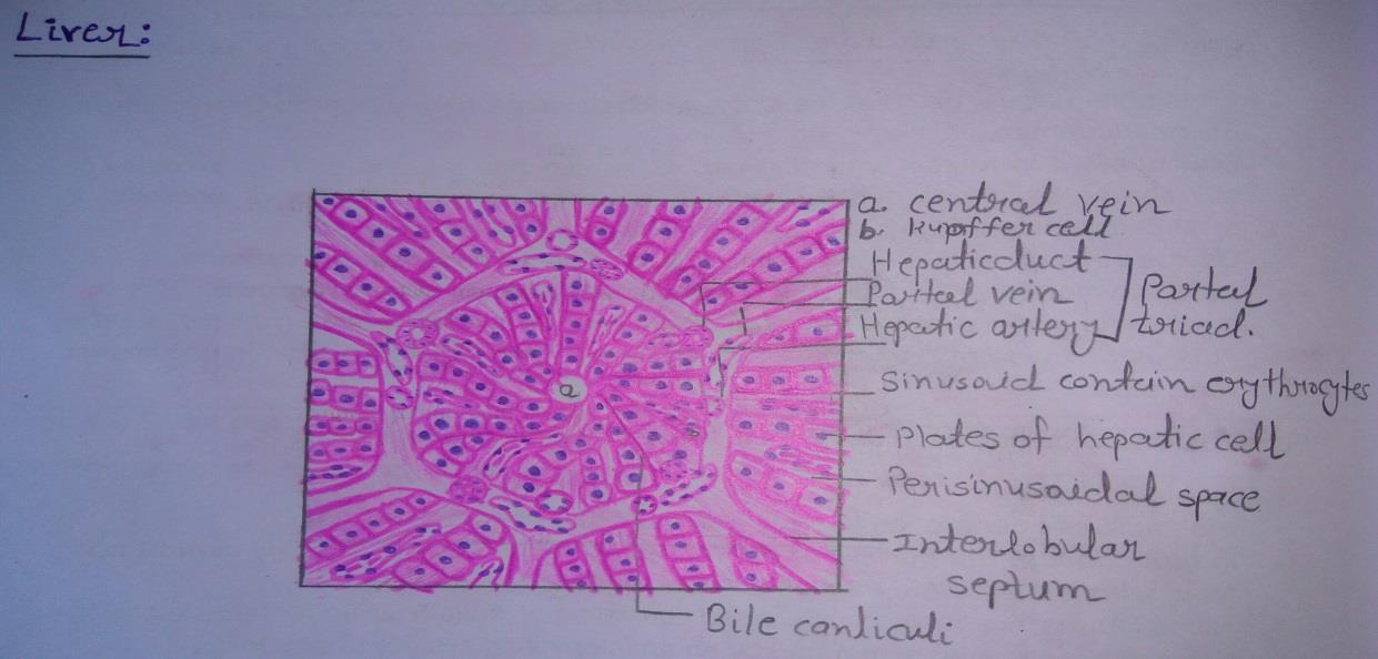
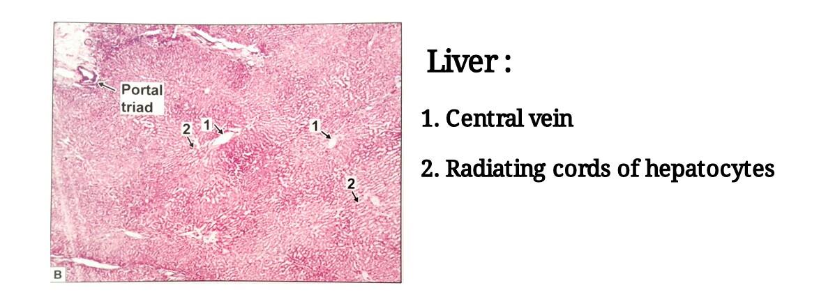
Slide 15: LIVER
DRAWING:
IDEAL SLIDE:
IDENTIFICATION POINTS:
| 1. The view of the Liver shows many hexagonal areas called hepatic lobules. The lobules are partially separated by connective tissue. |
| 2. Each lobule has a small round space in the center. This is the central vein. |
| 3. A number of broad irregular cords of cells seem to pass from this vein to the periphery of the lobule. These cords are made up of polygonal liver cells- hepatocytes. |
| 4. The cords are separated from each other by spaces called sinusoids. |
| 5. The sinusoids are lined by endothelial cells and Kupffer cells (macrophage cells). |
| 6. Along the periphery of the lobules there are angular intervals filled by connective tissue. |
| 7. Each such area contains a branch of the portal vein, a branch of the hepatic artery, and an interlobular bile duct. |
| 8. These three constitute portal triad. The identification of hepatic lobules and of portal triads is enough to recognize liver tissue. |
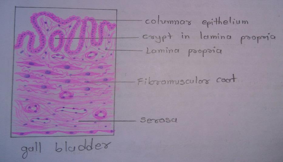
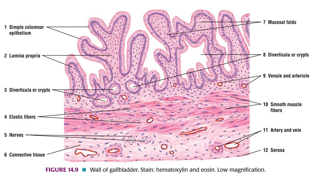
Slide 16: GALL BLADDER
DRAWING:
IDEAL SLIDE:
IDENTIFICATION POINTS:
| 1. The mucous membrane is lined by tall columnar cells with a striated border. |
| 2. The mucosa is highly folded and some of the folds might look like villi. |
| 3. Crypts may be found in lamina propria. |
| 4. Submucosa is absent. |
| 5. The muscle coat is poorly developed there being numerous connective tissue fibers amongst the muscle fibers. This is called as fibromuscular coat. |
| 6. A serous covering lined by flattened mesothelium is seen. |
| 7. Gall bladder can be differentiated from small intestine by- • Absence of villi • Absence of goblet cells • Absence of submucosa • Absence of proper muscularis externa |
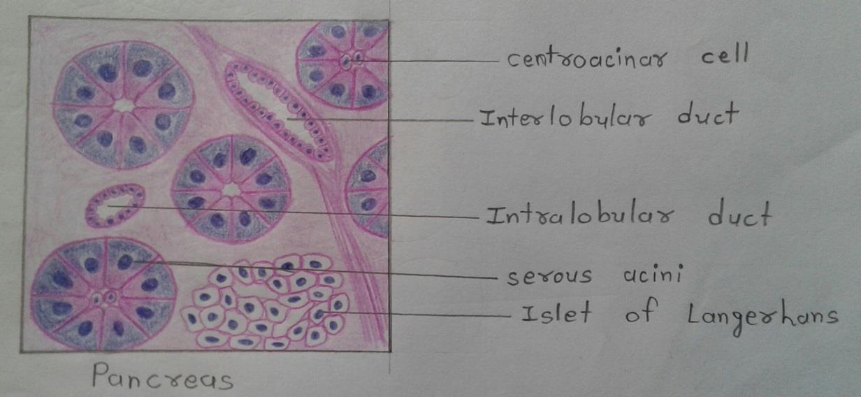
Slide 17: PANCREAS
DRAWING:
IDEAL SLIDE:
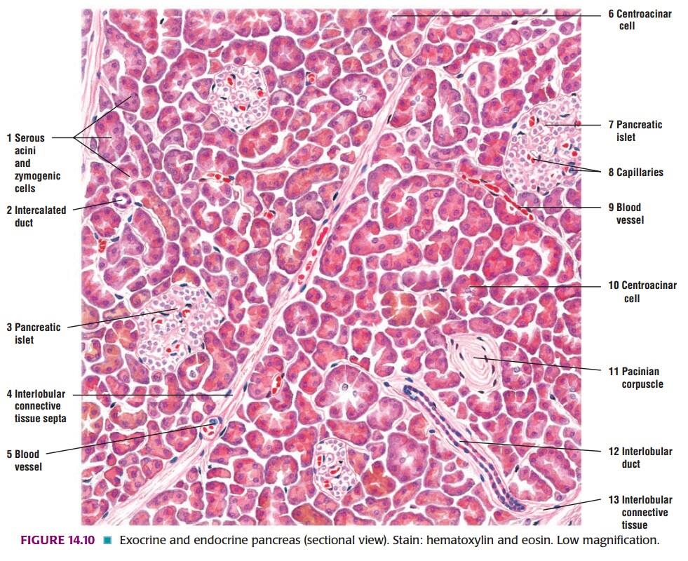
IDENTIFICATION POINTS:
| 1. This is a gland made up of serous acini. |
| 2. The lumen of the acinus is very small. |
| 3. In section stained with H & E, the cytoplasm of acinar cell is highly basophilic, particularly in the basal part. |
| 4. Numerous secretory granules can be demonstrated in the cytoplasm, especially in the apical part of the cell. These granules are eosinophilic. |
| 5. Some acini may show pale staining centroacinar cells in the center. |
| 6. Centroacinar cells really belong to the intercalated ducts. |
| 7. Amongst the acini some ducts are seen. |
| 8. The ducts have a distinct lumen, lined by cuboidal epithelium. |
| 9. At some places, the acini are separated by areas where we see aggregation of cells quite different from those of the acini. |
| 10. These aggregations form the pancreatic islets, pale staining cells arranged as groups, surrounded by blood vessels. |
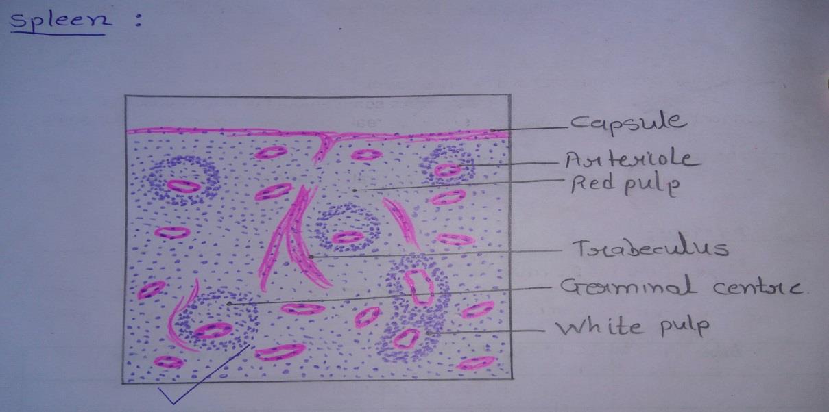
Slide 18: SPLEEN
DRAWING:
IDEAL SLIDE:
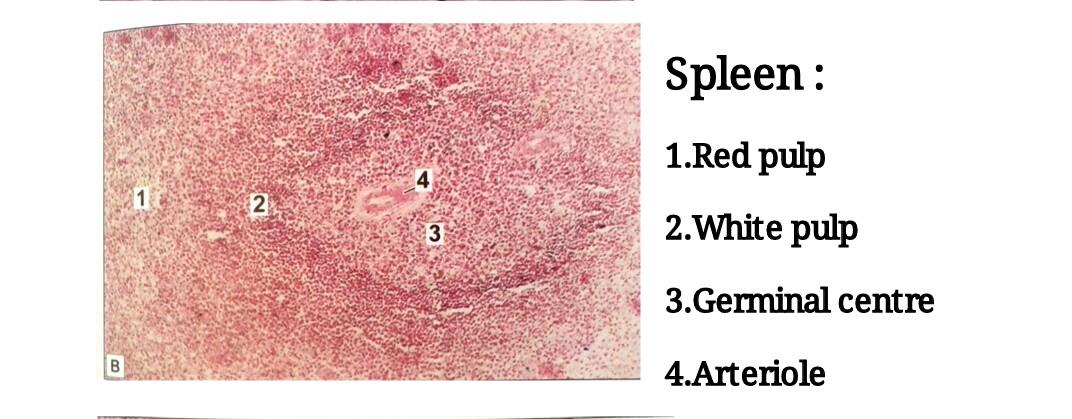
IDENTIFICATION POINTS:
| 1. The spleen is characterized by a thick capsule with trabeculae extending from it into the organ. |
| 2. The substance of the organ is divisible into the red pulp in which there are diffusely distributed lymphocytes and numerous sinusoids and the white pulp in which dense aggregation of lymphocytes are present. The latter are in the form of cords surrounding arterioles. |
| 3. When cut transversely, the cords resemble the lymphatic nodules of lymph nodes and like them, they have germinal centers surrounded by rings of densely packed lymphocytes. |
| 4. The nodules of the spleen are easily distinguished from those of lymph nodes because of the presence of arterioles in each nodule. |
| 5. This arteriole occupies an eccentric position in the nodules. |
| 6. More than one arteriole may be present in relation to one germinal center. |
Comments (1)