Urogenital (Systemic Histology 3)
6 years ago 21084
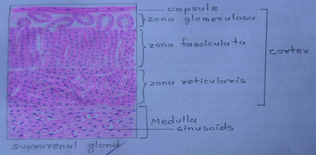
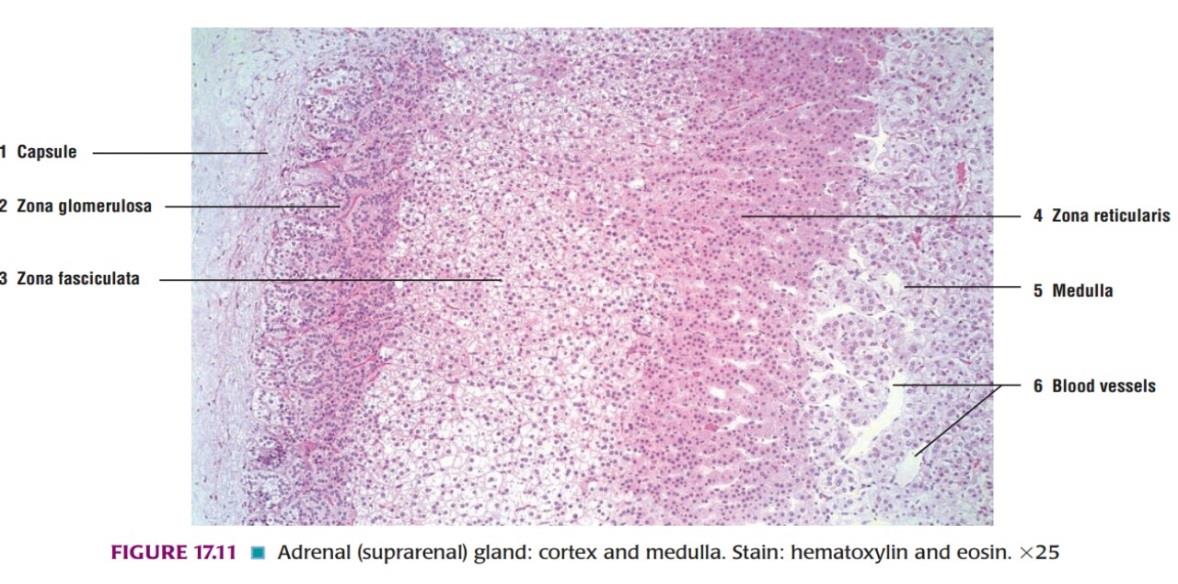
Slide 1: SUPRARENAL GLAND
DRAWING:
IDEAL SLIDE:
IDENTIFICATION POINTS:
| 1) In zona glomerulosa, the cells are arranged as inverted U-shaped formations. The cells of the zona glomerulosa are seen to be small, polyhedral, or columnar, with basophilic cytoplasm and deeply staining nuclei. |
| 2) In zona fasciculata, the cells are arranged in straight columns, two cells thick. Sinusoids intervene between the columns. The cells of the zona fasciculata are seen to be large, polyhedral, with basophilic cytoplasm and vesicular nuclei. |
| 3) The cells in zona reticularis are smaller and more acidophilic than the other two-layer. The zona reticularis is made up of cords of cells that branch and form a network. |
| 4) The medulla of the suprarenal gland is made up of chromaffin cells. The chromaffin cells are arranged in groups or columns which are separated by wide sinusoids. |
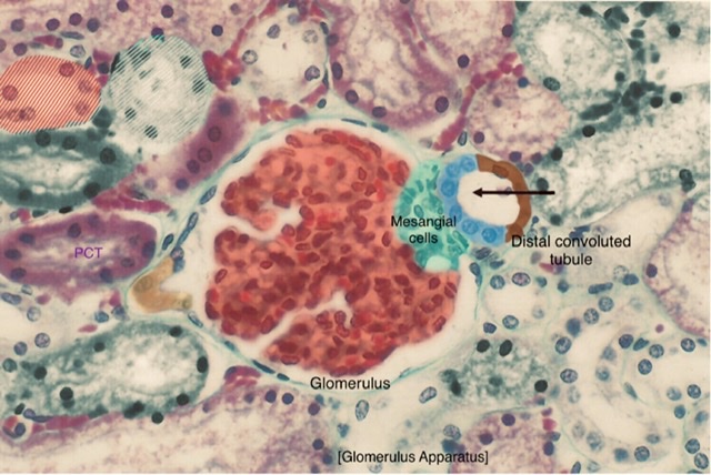
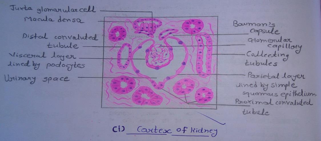
Slide 2: RENAL CORTEX
DRAWING:
IDEAL SLIDE:
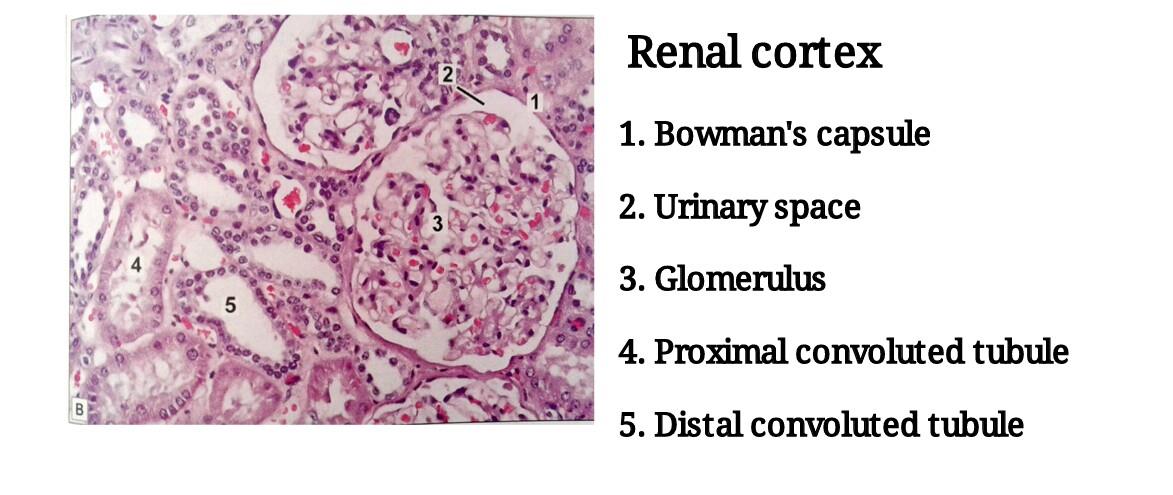
IDENTIFICATION POINTS:
| 1. The kidney is covered by a capsule. |
| 2. Deep to the capsule, there is the cortex. |
| 3. In the cortex, we see a circular structure called renal corpuscles surrounding which there are tubules cut in various shapes. |
| 4. The dark pink stained tubules are parts of the proximal convoluted tubules, there lumen is small and indistinct. It is lined by cuboidal epithelium with a brush border. |
| 5. Lighter staining tubules, each with a distinct lumen, are the distal convoluted tubules, they are lined by simple cuboidal epithelium. |
| 6. PCT are more in number than DCT. |
Slide 3: RENAL MEDULLA
DRAWING:

IDEAL SLIDE:
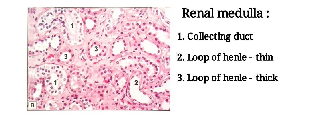
IDENTIFICATION POINTS:
| 1. A high power view of a part of the renal medulla shows a number of collecting ducts cut transversely or longitudinally. |
| 2. They are lined by a cuboidal epithelium, the cells of which stain lightly. Cell boundaries are usually distinct. The lumen of the tubules is also distinct. |
| 3. Sections of the thin segment of the loop of Henle are seen. They are lined by flattened cells, the walls being very similar in appearance to those of blood very capillaries. |
| 4. Sections through the thick segments of loops of Henle are seen. They are lined by cuboidal epithelium. |
Ø NOTE:
When we look at a section of the kidney, we see that most of the area is filled with a very large number of tubules. These are of various shapes and have different types of epithelial lining. This fact by itself suggests that the tissue is the kidney.
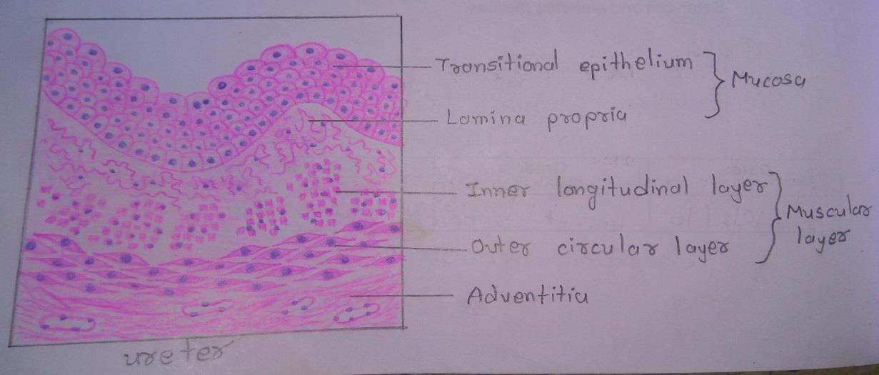
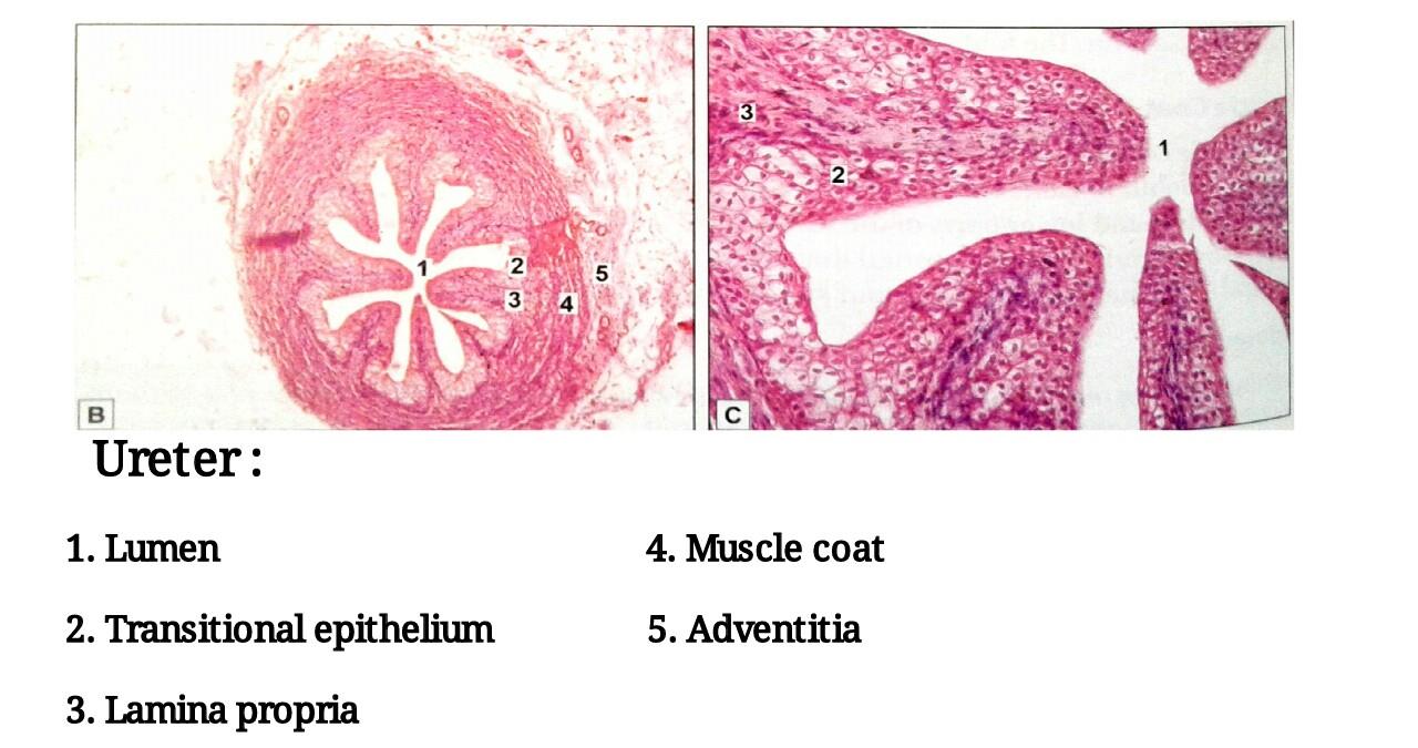
Slide 4: URETER
DRAWING:
IDEAL SLIDE:
IDENTIFICATION POINTS:
| 1. The Ureter can be recognized because it is tubular and its mucous membrane is lined by transitional epithelium. |
| 2. The epithelium rests on a lamina propria. |
| 3. The mucosa shows folds that give the lumen a star-shaped appearance. |
| 4. The muscle coat has an inner layer of longitudinal fibers and an outer layer of circular fibers. This arrangement is the reverse of that in the gut. |
| 5. The muscle coat is surrounded by connective tissue-adventitia in which blood vessels and fat cells are present. |
| 6. The ureter is differentiated from ductus deferens by a thin muscle coat and the presence of transitional epithelium. |
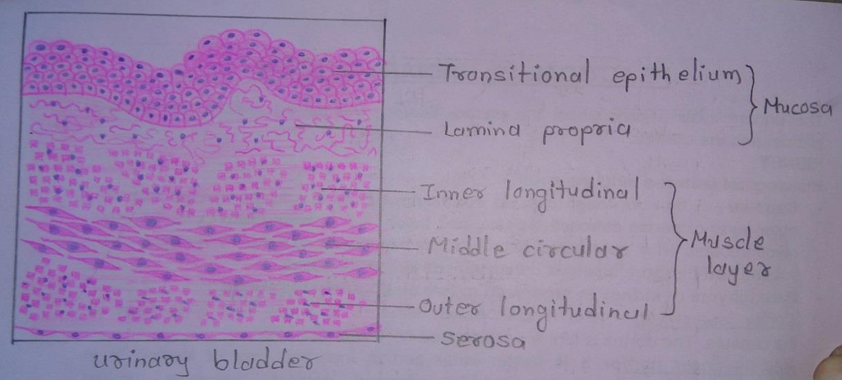
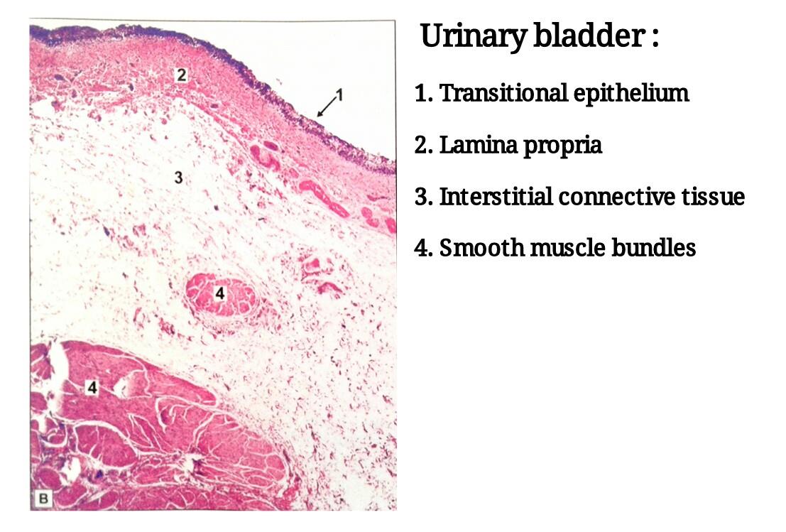
Slide 5: URINARY BLADDER
DRAWING:
IDEAL SLIDE:
IDENTIFICATION POINTS:
| 1. The mucous membrane is lined by transitional epithelium. There is no muscularis mucosae. |
| 2. In the empty bladder the mucous membrane is thrown into numerous folds that disappear when the bladder is distended. |
| 3. The muscle layer is thick. The smooth muscle in it forms a meshwork. Internally and externally the fibers tend to be longitudinal. In between them, there is a thicker layer of circular fibers. |
| 4. The distinct muscle layers may not be distinguishable. |
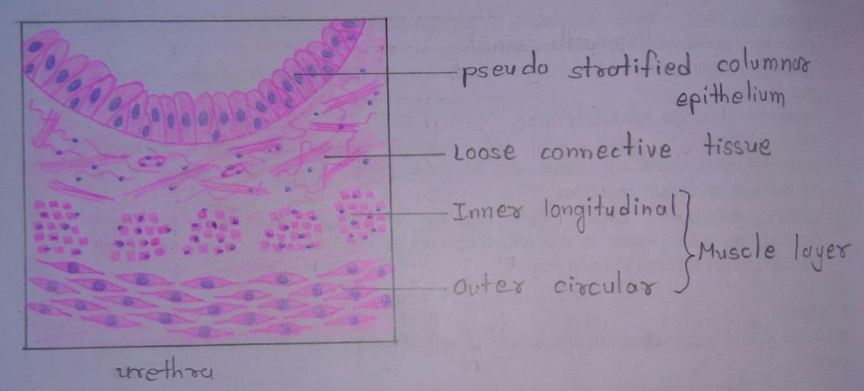
Slide 6: URETHRA
DRAWING:
IDEAL SLIDE:
IDENTIFICATION POINTS:
| 1. The mucous membrane consists of a pseudostratified columnar epithelium. A short part adjoining the urinary bladder is lined by transitional epithelium, while the part near the external orifice is lined by stratified squamous epithelium. |
| 2. The submucosa consists of loose connective tissue. |
| 3. The muscle coat consists of an inner longitudinal layer and an outer circular layer of smooth muscle. This coat is better defined in the female urethra. In the male urethra, it is well defined only in the membranous and prostatic parts. |
Male Reproductive System Histology
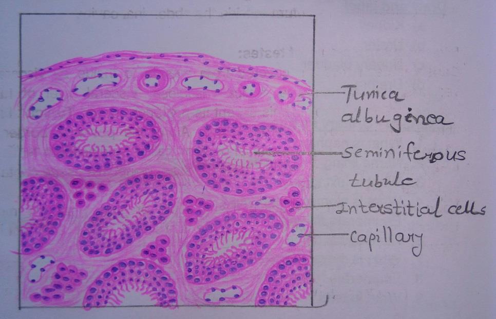
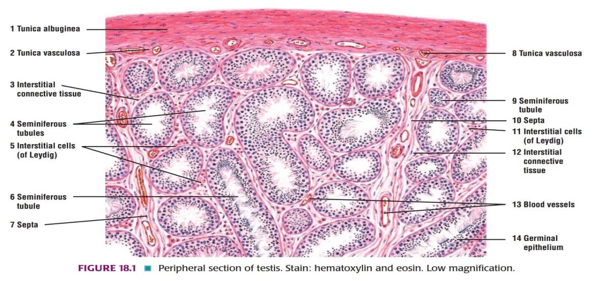
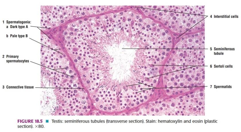
Slide 7: TESTIS
DRAWING:
IDEAL SLIDE:
IDENTIFICATION POINTS:
| 1. The testis has an outer fibrous layer, the tunica albuginea deep to which: ο A number of seminiferous tubules cut in various directions are seen. ο The tubules are separated by connective tissue, containing blood vessels and groups of interstitial cells of Leydig. ο Each seminiferous tubule is lined by several layers of cells. ο Cells are of two types: • Spermatogenic cells which produce spermatozoa, • Sustentacular (Sertoli) cells that have a supportive function. |
| 2. Details of cells lining a seminiferous tubule seen at a high magnification: i) The outermost row of nuclei belongs to sustentacular cells and to spermatogonia. ii) Passing inwards towards the centre of the tubule we have large darkly staining nuclei of spermatocytes, and many smaller nuclei of spermatids. iii) Towards the center of the tubule a number of developing spermatozoa are seen. |
| 3. In the practical class you may not be able to recognize these cells. Observe that the presence of many cells located at different levels gives the appearance of a stratified epithelium which is actually the spermatogonia at different stages of maturation. |
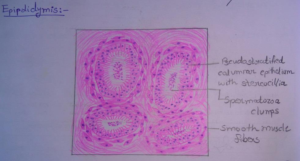
Slide 8: EPIDIDYMIS
DRAWING:
IDEAL SLIDE:
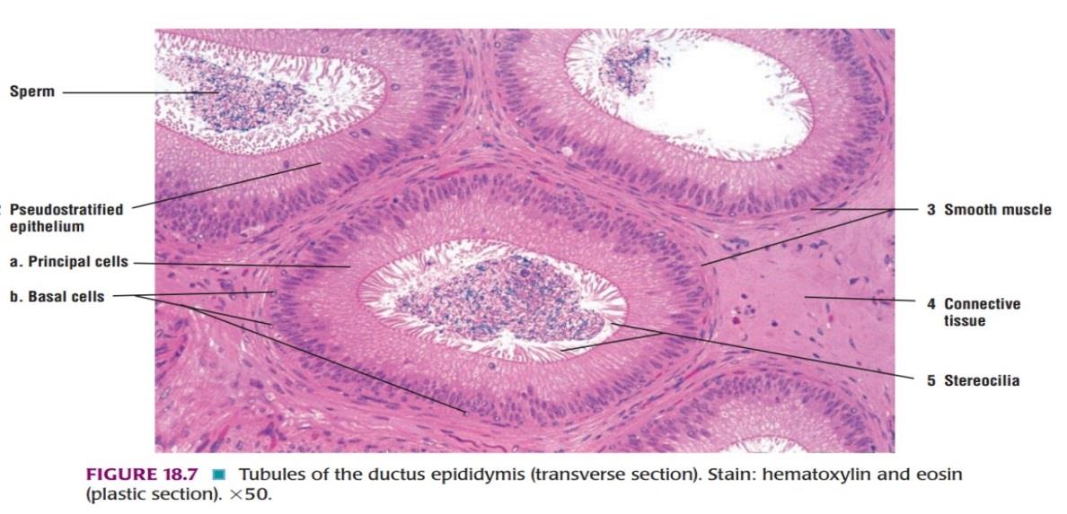
IDENTIFICATION POINTS:
| 1. The duct is lined by pseudostratified columnar epithelium which is made of 2 types of cells tall columnar cells, and shorter basal cells that do not reach the lumen. |
| 2. The luminal surface of each columnar cell bears non-motile projections that resemble cilia. They do not have the structure of true cilia. |
| 3. The basal cells are precursors of the tall cells. |
| 4. Beneath the epithelium there is a layer of circularly arranged smooth muscle fibres. This muscle layer increases in thickness gradually from head to tail. |
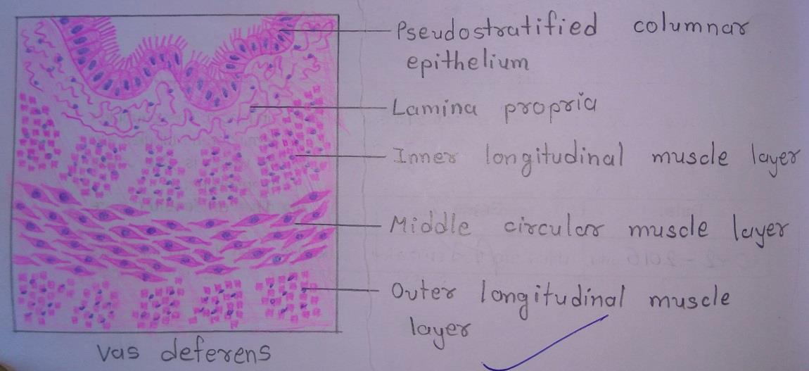
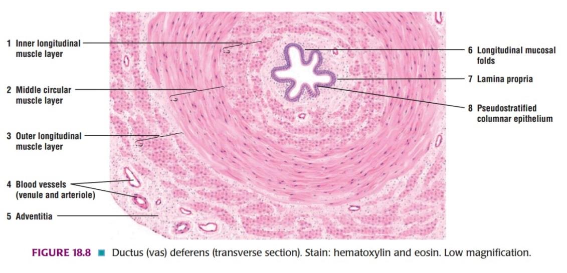
Slide 9: DUCTUS DEFERENS
DRAWING:
IDEAL SLIDE:
IDENTIFICATION POINTS:
| 1. The mucous membrane shows a number of longitudinal folds so that lumen appears to be stellate in section. |
| 2. The lining epithelium is simple columnar but becomes pseudostratified columnar in the distal part of the duct. |
| 3. The epithelium is supported by a lamina propria. |
| 4. The muscle coat is very thick and consists of smooth muscle. It is arranged in the form of an inner circular layer and outer longitudinal layer. |
| 5. The fibroelastic connective tissue forms the adventitial layer containing blood vessels and nerves. |
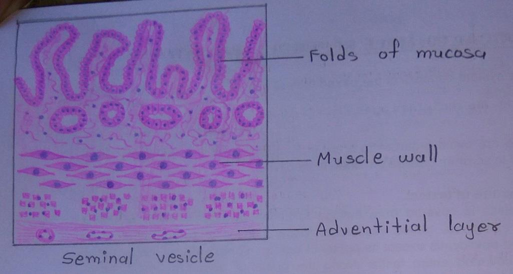
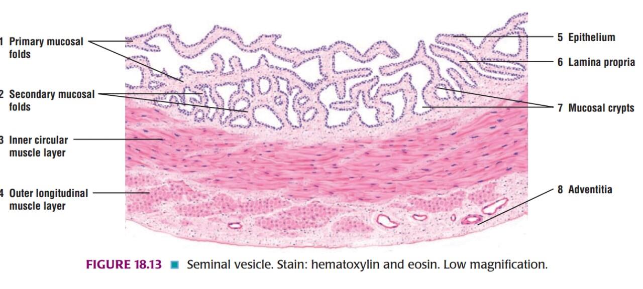
Slide 10: SEMINAL VESICLE
DRAWING:
IDEAL SLIDE:
IDENTIFICATION POINTS:
| 1. The mucous lining is thrown into numerous thin folds that branch and anastomose. The lining epithelium is simple columnar, or pseudostratified. Goblet cells are present in the epithelium. |
| 2. The seminal vesicles consists of a thin intermediate layer of smooth muscles. The muscle layer contains outer longitudinal and inner circular fibres. |
| 3. The outer covering of loose connective tissue forms the adventitial layer containing blood vessels and nerves. |
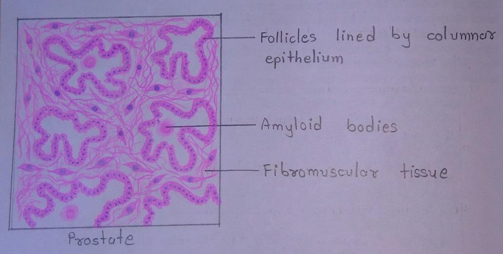
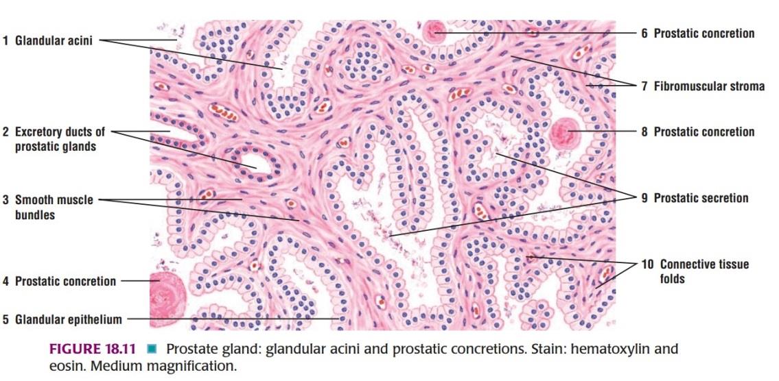
Slide 11: PROSTATE
DRAWING:
IDEAL SLIDE:
IDENTIFICATION POINTS:
| 1. The prostate consists of glandular tissue embedded in prominent fibromuscular stroma. |
| 2. The glandular tissue is in the form of follicles with serrated edges. They are lined by columnar epithelium. The lumen may contain amyloid bodies. |
| 3. The amyloid bodies or corpora amylacea are more abundant in older individuals. These consist of condensed glycoprotein. |
| 4. The follicles are separated by broad bands of fibromuscular tissue. |
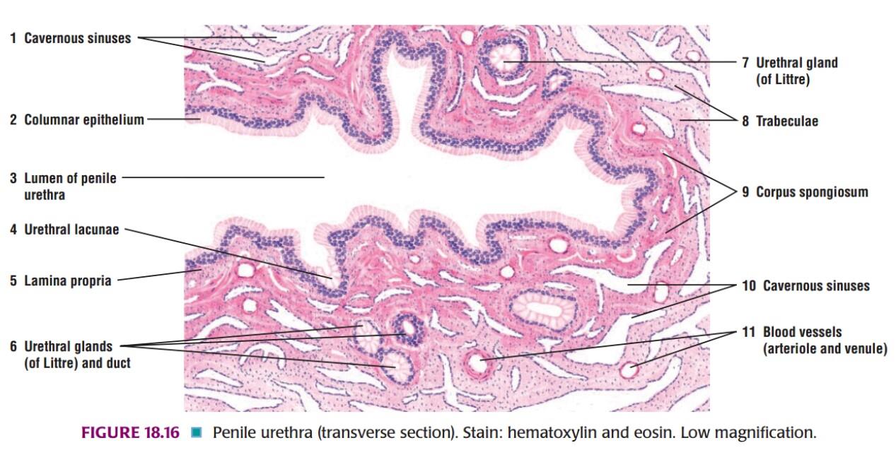
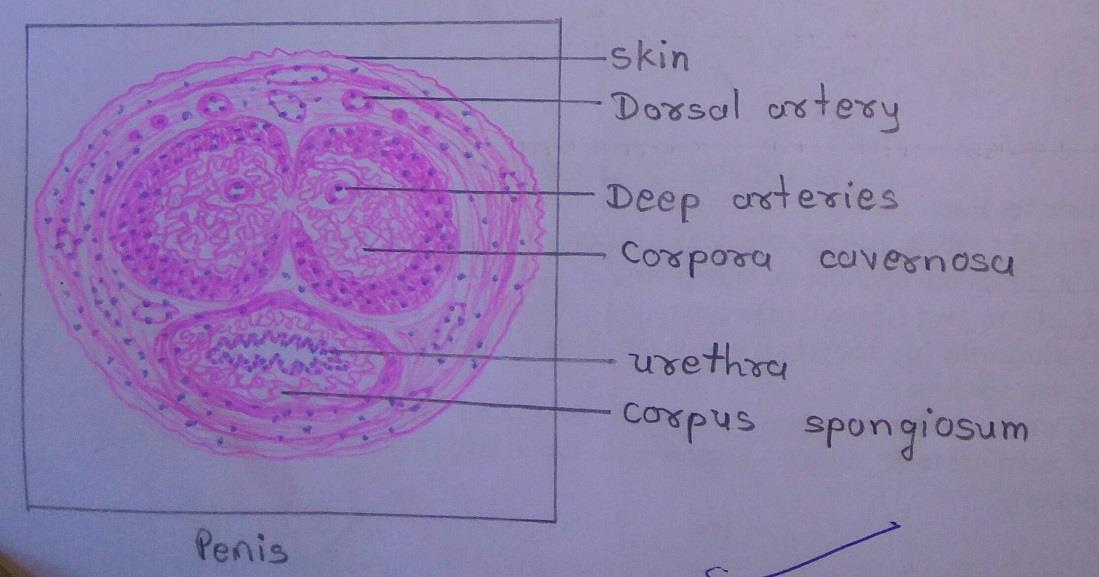
Slide 12: PENIS
DRAWING:
IDEAL SLIDE:
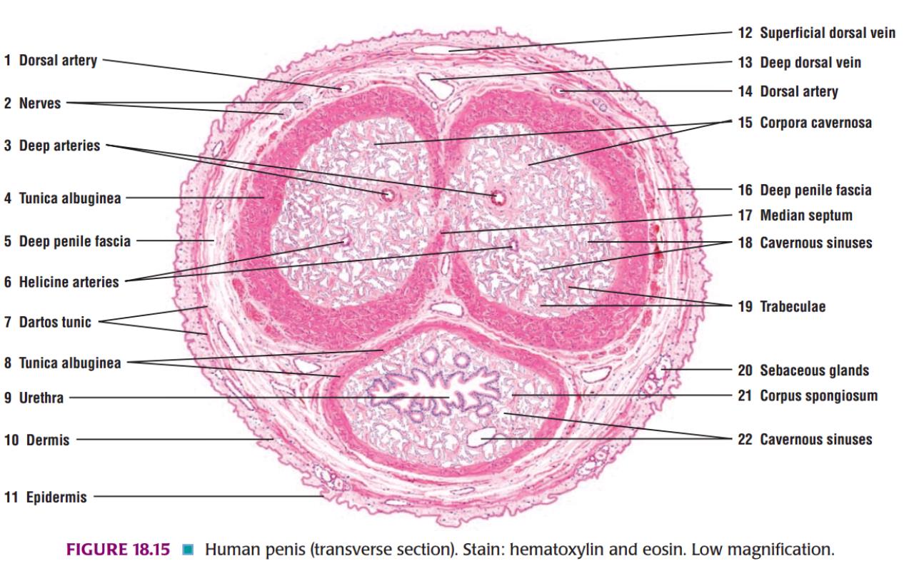
IDENTIFICATION POINTS:
| 1. The penis is covered all round by thin skin that is attached loosely to underlying tissue. |
| 2. The substance of the penis is made up of three masses of erectile tissue. The dorsal masses are the right and left corpora cavernosa, while the ventral mass is the corpus spongiosum. |
| 3. The corpus spongiosum is traversed by the penile urethra throughout its length. |
| 4. The tip of the urethra at the glans penis is lined by stratified squamous non-keratinized epithelium. |
| 5. Many small mucous glands of Littre are scattered along the length of the urethra that secrete mucus. |
Female Reproductive Histology
Slide 13: OVARY
DRAWING:
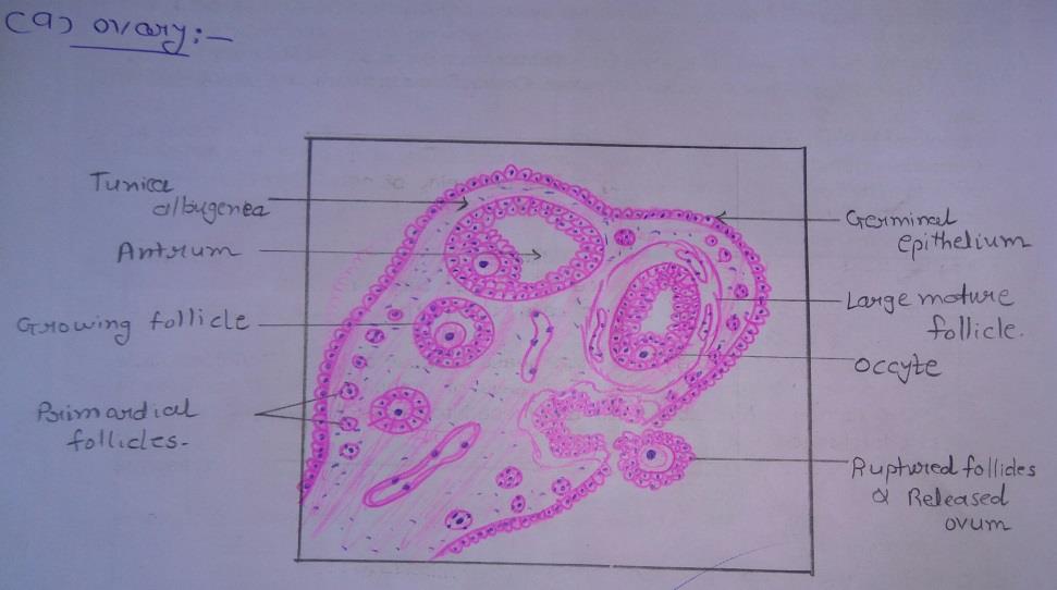
IDEAL SLIDE:
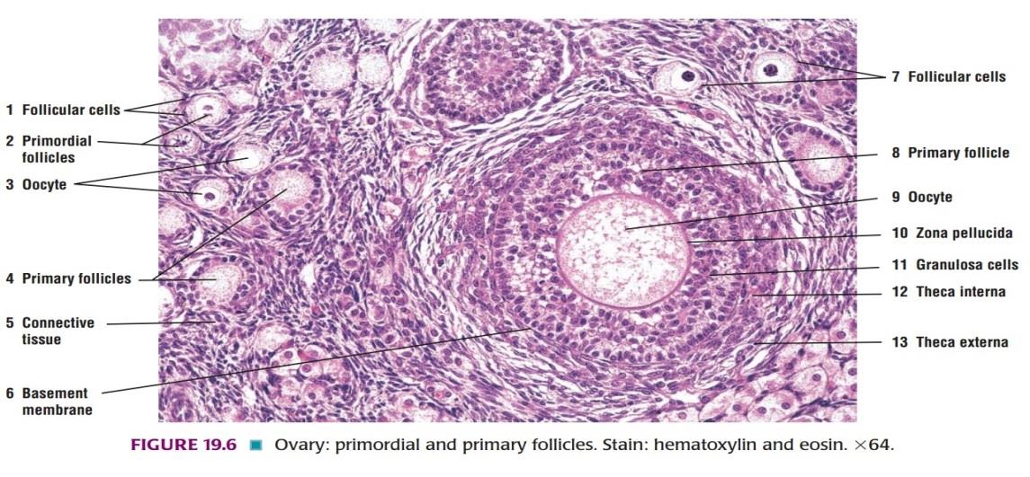
IDENTIFICATION POINTS:
| 1. The surface is covered by a cuboidal epithelium. Deep to the epithelium, there is a layer of connective tissue that constitutes the tunica albuginea. |
| 2. The substance of the ovary has an outer cortex in which follicles of various sizes are present; and an inner medulla consisting of connective tissue containing numerous blood vessels. |
| 3. Just deep to the tunica albuginea many primordial follicles each of which contains a developing ovum surrounded by flattened follicular cells are present. |
| 4. Large follicles have a follicular cavity surrounded by several layers of follicular cells. |
| 5. The cells surrounding the ovum constitute the cumulus oophorous. |
| 6. The follicle is surrounded by a condensation of connective tissue which forms a capsule for it. |
| 7. The capsule consists of an inner cellular part, and an outer fibrous part collectively called as theca folliculi. The follicle is surrounded by a stroma made up of reticular fibres and fusiform cells. |
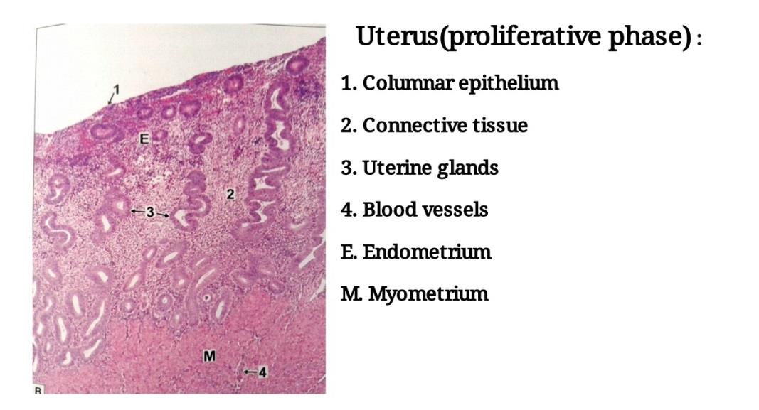
Slide 14: UTERUS IN PROLIFERATIVE PHASE
DRAWING:
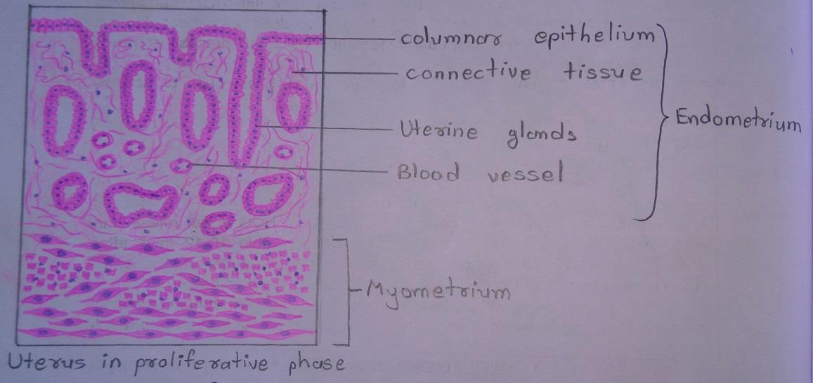
IDEAL SLIDE:
IDENTIFICATION POINTS:
| 1. The wall of the uterus consists of a mucous membrane (called the endometrium) and a very thick layer of muscle (the myometrium). The thickness of the muscle layer helps to identify the uterus easily. |
| 2. The endometrium has a lining of columnar epithelium that rests on a stroma of connective tissue. |
| 3. Numerous tubular uterine glands dip into the stroma. |
| 4. The appearance of the endometrium varies considerably depending upon the phase of the menstrual cycle • The endometrium is thin and progressively increases in thickness. • The uterine glands are straight and tubular in this phase. |
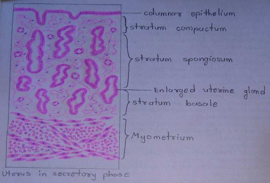
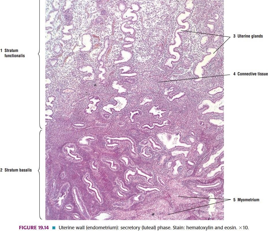
Slide 15: UTERUS IN SECRETORY PHASE
DRAWING:
IDEAL SLIDE:
IDENTIFICATION POINTS:
| 1. In the secretory phase: Ξ The thickness of the endometrium is much increased. Ξ The uterine glands elongate, become dilated, and tortuous as a result of which they have saw-toothed margins in sections. Ξ Blood vessels extend in the upper portion of endometrium. |
| 2. In this phase the appearance of the endometrium becomes so distinctive that the uterus cannot be confused with any other organ. |
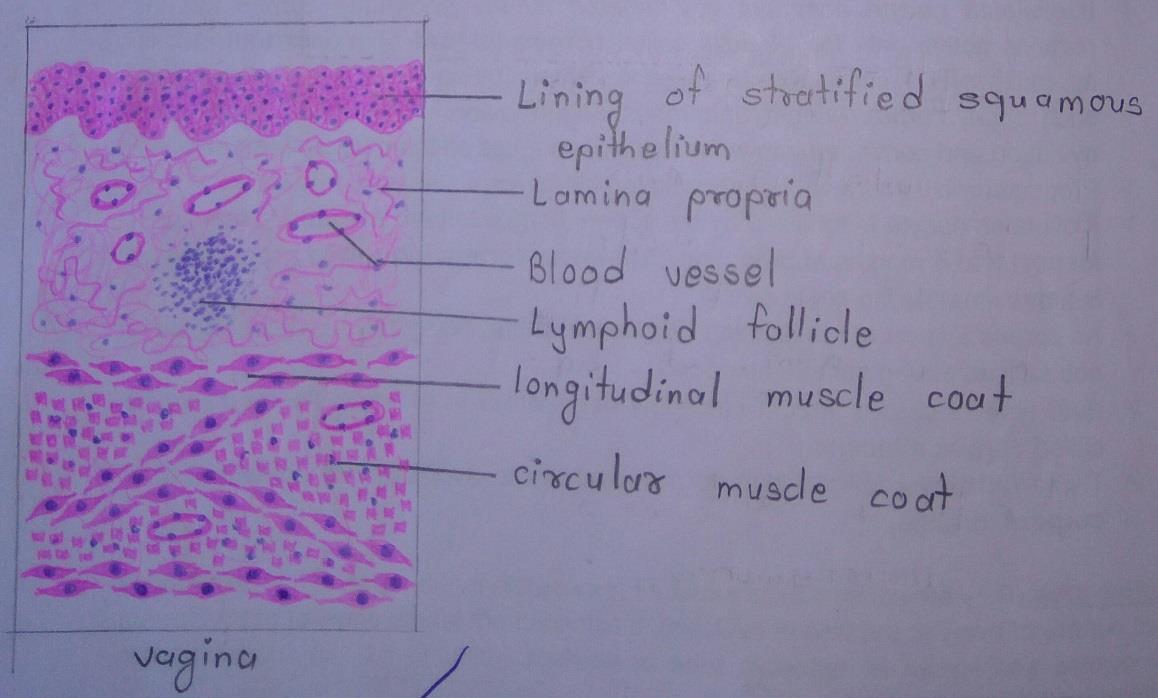
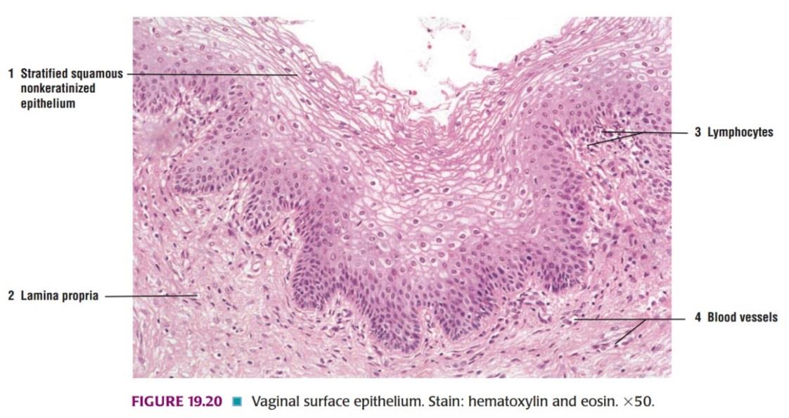
Slide 16: VAGINA
DRAWING:
IDEAL SLIDE:
IDENTIFICATION POINTS:
| 1. The mucous membrane shows numerous longitudinal folds and is firmly fixed to the underlying muscle layer. It is lined by non-keratinized stratified squamous epithelium. |
| 2. No glands are seen in the mucosa. The mucosa of the vagina is rich in glycogen and hence the cells are pale stained which distinguishes it from the esophagus. |
| 3. The muscle coat is made up of an outer layer of longitudinal fibers, and a much thinner inner layer of circular fibers. |
| 4. The muscle wall is surrounded by an adventitia made up of fibrous tissue containing many elastic fibers. |
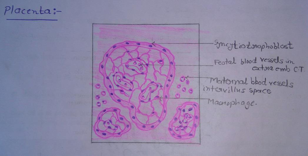
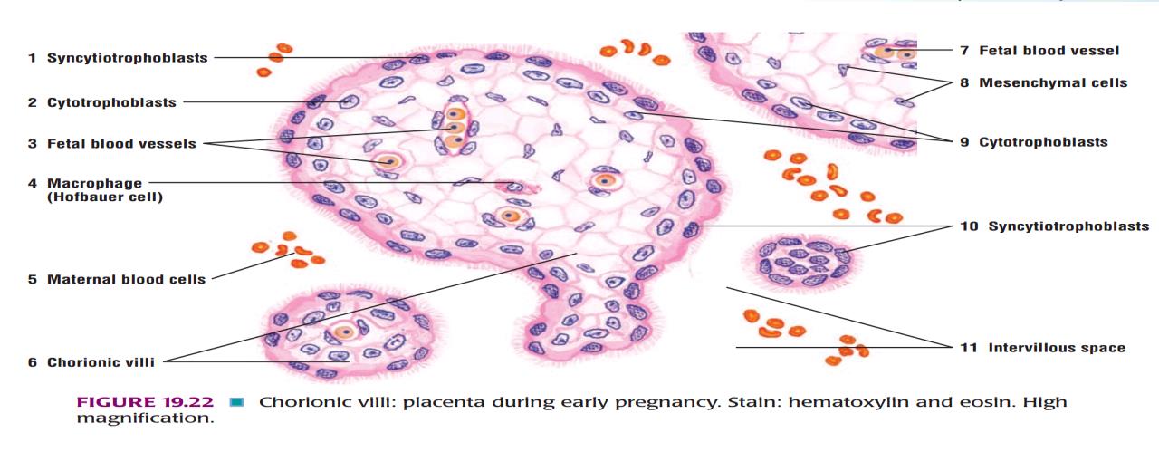
Slide 17: PLACENTA
DRAWING:
IDEAL SLIDE:
IDENTIFICATION POINTS:
| 1. A slide of the placenta shows numerous chorionic villi. |
| 2. A villus is lined with inner cytotrophoblasts and outer syncytiotrophoblasts. |
| 3. Cytotrophoblasts are cuboidal in shape. |
| 4. Syncytiotrophoblasts layer is consists of multinucleated cytoplasm with indistinct cell margins. |
| 5. Core of villi contains umbilical blood capillaries embedded in thin layers of foetal connective tissue. |
| 6. Cross-sections of villi are surrounded by maternal blood. |
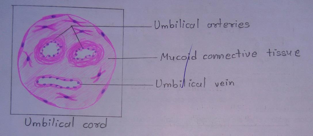
Slide 18: UMBILICAL CORD
DRAWING:
IDENTIFICATION POINTS:
| 1. It contains two umbilical arteries, which carries deoxygenated blood from fetus to placenta. |
| 2. It also contains one umbilical vein, which carries oxygenated blood from the placenta to the fetus. |
| 3. Amniotic membrane lined by flattened epithelial cells. |
| 4. Deep to amniotic membrane, mucoid connective tissue [Wharton`s Jelly] is present which contains fibroblasts, collagen fibers, and ground substance. |
Comments (0)