Muscle Physiology (Viva)
6 years ago 22245
Q.1 How are the muscles classified?
By three methods:
Q.2 Which are the striated muscles?
Skeletal muscles and cardiac muscles are striated muscles.
Q.3 What is the difference between the skeletal, cardiac, and smooth muscles?
The difference between skeletal, cardiac, and smooth muscles is shown in the table.
| Features | Skeletal muscle | Cardiac muscle | Smooth muscle |
| Location | In association with bones | In the heart | In the visceral organs |
| Shape | Cylindrical and unbranched | Branched | Spindle-shaped, unbranched |
| Length | 1-4 cm | 80-100 μ | 50-200 μ |
| Diameter | 10-100 μ | 15-20 μ | 2-5 μ |
| No. of nucleus | More than one | One | One |
| Cross striations | Present | Present | Absent |
| Myofibrils | Present | Present | Absent |
| Sarcomere | Present | Present | Absent |
| Troponin | Present | Present | Absent |
| Sarcotubular system | Well developed | Well developed | Poorly developed |
| T tubules | Long and thin | Short and broad | Absent |
| Depolarization | Upon stimulation | Spontaneous | Spontaneous |
| Fatigue | Possible | Not possible | Not possible |
| Summation | Possible | Not possible | Possible |
| Tetanus | Possible | Not possible | Possible |
| Resting membrane potential | Stable | Stable | Unstable |
| For trigger of contraction, calcium binds with | Troponin | Troponin | Calmodulin |
| Source of calcium | Sarcoplasmic reticulum | Sarcoplasmic reticulum | Extracellular |
| Speed of contraction | Fast | Intermediate | Slow |
| Neuromuscular junction | Well defined | Not well defined | Not well defined |
| Action | Voluntary action | Involuntary action | Involuntary action |
| Control | Only neurogenic | Myogenic | Neurogenic and myogenic |
| Nerve supply | Somatic nerves | Autonomic nerves | Autonomic nerves |
Q.4 What is the nerve supply of different types of muscles?
Skeletal muscle is supplied by somatic nerves. Cardiac and smooth muscles are supplied by autonomic nerve fibers.
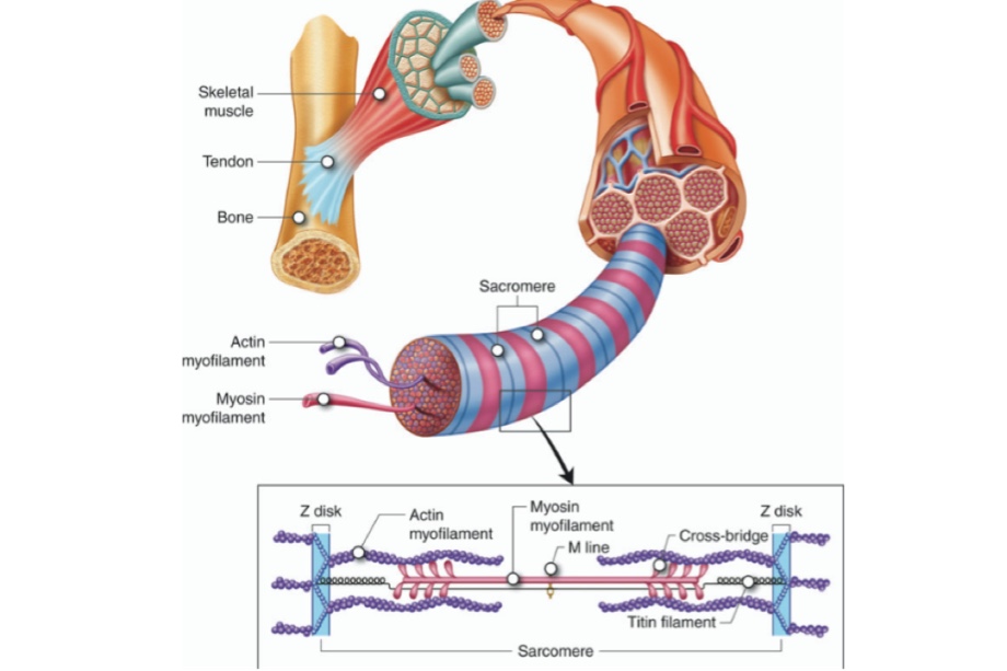
Q.5 What are myofibrils?
Myofibrils are the thin parallel filaments present in the sarcoplasm of the muscle fiber.
Q.6 What is sarcomere?
The structural and functional unit of skeletal muscle is known as sarcomere. It extends between two ‘Z’ lines.
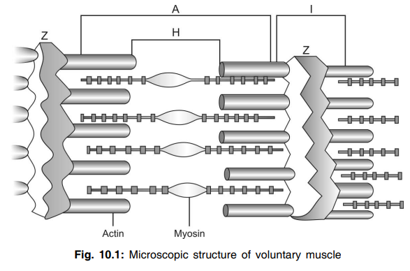
Q.7 Discuss in short microscopic structure of voluntary muscle cell.
A muscle cell consists of alternate transverse dark (anisotropic) A-band, and light (isotropic) I-band. A-band has in its center a region of low refractive index (H-band or Hensen line), and I-band a line of high refractive index (Z-line or Dobie line).
Q.8 What is ‘A’ band in the muscle? Why is it called so?
‘A’ band is the dark band present in the myofibrils of the muscle. It is anisotropic to polarized light; i.e., if polarized light is passed through this area of the muscle, the light rays are refracted in different directions. So this band is called ‘A’ band.
Q.9 What is ‘I’ band in the muscle? Why is it called so?
‘I’ band is the light band present in the myofibrils of the muscle. It is isotropic to polarized light, i.e. when polarized light is passed through this area of the muscle, all the light rays are refracted at the same angle. So this band is called ‘I’ band.
Q.10 What are the myofilaments?
Myofilaments are the thread-like protein filaments present in the sarcomere. Myofilaments are of two types, actin filaments and myosin filaments.
Q.11. What are the myofilaments present in the ‘A’ band?
Myosin filaments and part of actin filaments.
Q.12 What are the myofilaments present in ‘I’ band?
Actin filaments.
Q.13 Explain the features and situation of myofilaments briefly.
Actin filaments are thin filaments with a diameter of 20 Å and extend from either side of the ‘Z’ lines, run across the ‘I’ band, and enter into the ‘A’ band up to the ‘H’ zone. Myosin filaments are thick filaments with a diameter of 115 Å and are situated in the center of the ‘A’ band.
Q.14 What are the components of actin and myosin filaments?
The actin filament consists of three types of proteins called actin, tropomyosin, and troponin. The myosin filament consists of myosin molecules.
Q.15 What are the contractile elements of the skeletal muscle?
The contractile elements of the skeletal muscle are the muscle proteins namely myosin, actin, tropomyosin and troponin.
Q.16 What is ‘H’ zone? And what is ‘M’ line?
‘H’ zone is a light area in the middle of the ‘A’ band. ‘M’ band is the middle part of myosin filaments situated in the middle of the ‘H’ zone.
Q.17 What is sarcotubular system? What are its components?
Sarcotubular system is a system of membranous tubular structures present in the skeletal muscle fiber. The components of this system are ‘T’ tubules (transverse tubules) and ‘L’ tubules (longitudinal tubules). ‘L’ tubule is otherwise called sarcoplasmic reticulum.
Q.18 What is the functional importance of sarcotubular system?
The ‘T’ tubules are responsible for the rapid transmission of the action potential through the muscle fiber. The “L” tubules store a large quantity of calcium ions.
Q.19 What are the organic substances present in skeletal muscle?
Q.20 Name the properties of skeletal muscle.
The properties of skeletal muscle are excitability, contractility, and muscle tone.
Q.21 Define excitability.
The response of the living tissue to a stimulus in the form of physicochemical change is known as excitability.
Q.22 What is action potential?
Conduction of nerve signal by depolarization which changes the normal resting negative potential to positive potential followed by repolarization back to the normal negative membrane potential is called Action Potential.
Q.23 Define stimulus. What are the types of stimulus?
Stimulus is an agent or influence that brings about the response in an excitable tissue.
Stimulus is of four types – mechanical, electrical, thermal, and chemical stimulus.
Q.24 Name the qualities of a stimulus.
Q.25 What is the strength-duration curve? What is its other name?
The strength-duration curve is the curve that demonstrates the relationship between the strength and the duration of the stimulus. It is also known as the excitability curve.
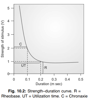
Q.26 What is rheobase?
Rheobase is the minimum strength of the stimulus that is required to excite the tissue.
Q.27 What is utilization time?
Utilization time is the minimum time required to excite the tissue when a stimulus with rheobasic strength (threshold strength of stimulus) is applied.
Q.28 What is chronaxie?
Chronaxie is the minimum time required to excite the tissue when a stimulus with double the rheobasic strength is applied.
Q.29 What is the importance of chronaxie?
Chronaxie helps to determine the excitability of the tissue. Longer the chronaxie, the lesser is the excitability.
Q.30 Name some conditions when chronaxie increases.
Q.31 What are the types of muscular contractions?
Q.32 Define isotonic contraction and give example.
Isotonic contraction is the type of contraction in which the tension remains the same and change occurs only in the length of the muscle fibers. An example is the contraction of the biceps muscle during simple flexion of the arm.
Q.33 Define isometric contraction and give example.
Isometric contraction is the type of contraction in which the length of the muscle fibers remains the same and change occurs only in the tension. An example is the contraction of arm muscles while pulling any heavy object.
Q.34 What is preload?
It is the load on a muscle in a relaxed state.
Q.35 What is afterload?
It is the load that the muscle must generate to overcome the higher pressure.
Q.36 What are the different periods in a simple muscle twitch?
Q.37 Give the normal duration of different periods of a simple muscle twitch.
Latent period = 0.01 sec
Contraction period = 0.04 sec
Relaxation period = 0.05 sec
Total twitch period = 0.10 sec
Q.38 Why is the contraction period shorter than the relaxation period?
The contraction period is shorter than the relaxation period because the contraction is an active process and relaxation is a passive process.
Q.39 Define latent period.
The latent period is defined as the time interval between the point of stimulus and point of contraction.
Q.40 What are the causes for the latent period?
Q.41 Name some conditions when the latent period is prolonged.
Q.42 When does the latent period decrease?
The latent period decreases when temperature is increased.
Q.43 Classify the skeletal muscles depending upon the contraction time. Give examples.
Slow or red muscles, which have longer contraction time. Examples: back muscles
Fast or pale muscles that have shorter contraction time. Examples: hand muscles and ocular muscles.
Q.44 What are the differences between red and white muscle fibers?
| Red (Slow muscle) | Pale (Fast muscle) |
| Type I fibers are more | Type II fibers are more |
| Myoglobin content is high. So. it is red | Myoglobin content is less. So, it is pale |
| Sarcoplasmic reticulum is less extensive | Sarcoplasmic reticulum is more extensive |
| Blood vessels are more extensive | Blood vessels are less extensive |
| Mitochondria are more in number | Mitochondria are less in number |
| Response is slow with long latent period | Response is rapid with short latent period |
| Contraction is less powerful | Contraction is more powerful |
| This muscle is involved in prolonged and continued activity as it undergoes sustained contraction | This muscle is not involved in prolonged and continued activity as it relaxes immediately. |
| Fatigue occurs slowly | Fatigue occurs quickly |
| Depends upon cellular respiration for ATP production | Depends upon glycolysis for ATP production |
Q.45 What are the factors affecting the force of contraction of the muscle within physiological limits?
Q.46 Classify the stimulus depending upon the strength.
Q.47 What is the threshold stimulus?
Threshold or minimal stimulus is the stimulus with minimum strength required to cause minimum response in the tissues.
Q.48 What are the effects of two successive stimuli on muscle?
Q.49 What is a beneficial effect?
When two stimuli are applied to a muscle one after another in such a way that the second stimulus falls after the relaxation period of the first twitch, two separate contractions are recorded and the force of the second contraction is greater than that of the first contraction. This is known as a beneficial effect.
Q.50 What is the cause for beneficial effects?
An increase in the temperature during the first contraction decreases the viscosity of muscle. So, the force of the second contraction is more.
Q.51 What is superposition?
While applying two successive stimuli, if the second stimulus falls during relaxation of the first twitch, the first curve is superimposed by the second curve. This is called superposition or incomplete summation.
Q.52 What is summation?
When two stimuli are applied one after another and if the second stimulus falls during the contraction period or second half of the latent period, two contractions are summed up, giving a single contraction which is bigger and broader than a simple muscle curve. This is known as summation or complete summation.
Q.53 Define fatigue.
The decrease in the response of the muscle due to repeated stimuli is known as fatigue.
Q.54 What are the causes of fatigue?
Q.55 Mention the order of site (seat) of fatigue in the intact body.
First site of fatigue: Cerebral cortex (Betz cells)
Second site of: Motor neuron in fatigue spinal cord
Third site of: Neuromuscular fatigue junction
Fourth site of fatigue: Muscle.
Q.56 How to prove that the neuromuscular junction is the first site of fatigue in the frog’s muscle nerve preparation?
In the isolated muscle nerve preparation, the nerve is stimulated continuously and the curves are recorded till the fatigue occurs, i.e. till the muscle fails to respond to the stimulus. Then, immediately the muscle is stimulated directly. A response is noticed in the form of a curve. This shows that the muscle is not yet fatigued. The nerve cannot be fatigued. So, the site where fatigue must have occurred is the neuromuscular junction.
Q.57 Is fatigue a reversible or irreversible phenomenon?
Fatigue is a reversible phenomenon.
Q.58 What are the causes of recovery from fatigue?
Q.59 What is tetanus?
Summation or complete fusion of muscular contractions due to repeated stimuli is known as tetanus. Tetanus is defined as the sustained contraction of muscle due to repeated stimuli.
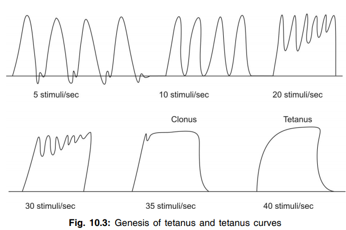
Q.60 What is clonus?
When the frequency of stimuli is not sufficient to cause tetanus, the fusion of contraction is not complete. This is known as clonus or incomplete tetanus.
Q.61 What is the frequency of stimuli to cause tetanus and clonus?
Frog muscle:
Frequency of stimuli to cause tetanus = 40/sec
Frequency of stimuli to cause clonus = 35/sec
Human muscle: Frequency of stimuli to cause
tetanus = 60/sec Frequency of stimuli to cause
clonus = 55/sec
Q.62 What is pathological tetanus?
Pathological tetanus is a disease caused by bacillus Clostridium tetani. It affects the nervous system and its common features are muscle spasm and paralysis.
Q.63 What is the effect of a moderate increase in temperature on the muscle? What are the causes of the effect?
Moderate increase in temperature to about 30 to 40° C, increases the force of contraction and decreases all the periods, i.e. the activity is accelerated.
Causes:
• Increase in excitability of the muscle
• Acceleration of chemical processes
• Decrease in the viscosity of the muscle.
Q.64 What is the effect of a decrease in temperature on the muscle? What are the causes of the effect?
Decrease in temperature to about 10° C, reduces the force of contraction and increases all the periods, i.e. the activity is slowed down
Causes:
Q.65 What is the effect of very high temperature on the muscle?
When the temperature increases above 60° C, heat rigor occurs.
Q.66 What is heat rigor? What is its cause?
Stiffening and shortening of the muscle fibers because of high temperature is called heat rigor. It is due to the coagulation of muscle proteins.
Q.67 Is heat rigor reversible?
Heat rigor is not reversible.
Q.68 What is cold rigor? Is it reversible?
Stiffening and shortening of the muscle fibers due to extreme cold is called cold rigor and it is reversible.
Q.69 What is calcium rigor? Is it reversible?
Rigor due to increased calcium content is known as calcium rigor. It is reversible.
Q.70 What is rigor mortis? What is the cause of it?
The rigidity that develops after death is called rigor mortis.
Cause:
After death, there is loss of ATP. Relaxation cannot occur because of lack of ATP and that is the cause of rigor mortis.
Q.71 What is freeload? Give an example.
Freeload or fore load is the load that acts on the muscle freely even before the onset of the contraction of the muscle.
Example: Filling water from a tap by holding the bucket in hand.
Q.72 State whether the muscle works better in after loaded condition or in free loaded condition. Why?
Muscle works better in free loaded condition than in the after loaded condition. Because, in free loaded condition the initial length of the muscle fibers increases even before the onset of muscular contraction. And according to Frank Starling’s law, the force of contraction of the muscle is directly proportional to the initial length of the muscle fiber within physiological limits.
Q.73 What is the optimum load?
Optimum load is the load at which the work done by the muscle is maximum.
Q.74 What is the refractory period?
The refractory period is the period at which the muscle does not show any response to a stimulus.
Q.75 What are the types of refractory period?
Absolute refractory period—the period during which the muscle does not show any response at all, whatever may be the strength of the stimulus
Relative refractory period—the period during which the muscle shows some response if the strength of stimulus is increased to maximum.
Q.76 What is the duration of absolute and relative refractory periods in skeletal muscle?
The absolute refractory period extends for 0.005 sec, i.e. during the first half of the latent period. The relative refractory period extends for 0.005 sec, i.e. during the second half of the latent period. Thus, the duration of the refractory period in skeletal muscle is 0.01 sec.
Q.77 What is the duration of absolute and relative refractory periods in cardiac muscle?
The absolute refractory period is 0.27 sec, i.e. it extends throughout the contraction period. The relative refractory period is 0.25 sec, i.e. it extends during the first half of the relaxation period. Thus, totally the refractory period in cardiac muscle extends for about 0.52 sec. It is very long compared to that of skeletal muscle.
Q.78 What is the significance of the long refractory period in cardiac muscle?
Because of the long refractory period, fatigue, tetanus, and complete summation cannot be produced in cardiac muscle.
Q.79 What is muscle tone?
The muscle fibers always maintain a state of slight contraction with a certain degree of vigor and tension. This is known as muscle tone or tonus.
Q.80 How is the tone maintained in skeletal and cardiac muscle?
Skeletal muscle: Maintenance of tone is neurogenic and it is under the influence of the gamma motor neuron system.
Cardiac muscle: Maintenance of tone is purely myogenic and it is by the muscle itself.
Q.81 Name the changes taking place during muscular contraction.
Q.82 What is resting membrane potential (RMP)?
The potential difference between inside and outside of the cell across the cell membrane under resting conditions is known as RMP. It is negative inside and positive outside.
Q.83 What are the mechanisms involved in the ionic basis of RMP?
Two transport mechanisms are involved in the ionic basis of RMP.
Q.84 How much is the RMP in skeletal muscle?
RMP in skeletal muscle is – 90 mV.
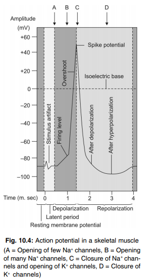
Q.85 What is action potential?
Series of electrical changes taking place in the cell when stimulated is known as an action potential.
Q.86 What are the properties of action potential?
Action potential:
Q.87 What are the phases of the action potential?
Q.88 What is depolarization?
When stimulated, the resting membrane potential is lost in the cell. The Interior of the cell becomes positive (up to +55 mV) and the exterior becomes negative. This is known as depolarization.
Q.89 What is the cause for depolarization?
Depolarization is due to the opening of sodium channels and the rush of sodium ions into the cell.
Q.90 Why the depolarization is short-lived?
Because of the rapid inactivation and closure of sodium channels.
Q.91 What is repolarization?
The restoration of negativity inside the cell and positivity outside is known as repolarization.
Q.92 What is the cause for repolarization?
Repolarization is due to the opening of potassium channels and the efflux of potassium ions from inside to outside the cell.
Q.93 What is the firing level?
When the cell is stimulated, depolarization starts slowly. After the initial slow depolarization up to – 15 mV, the rate of depolarization increases suddenly. The point at which the rate of depolarization increases is known as the firing level.
Q.94 What is spike potential?
During an action potential, the rapid depolarization and rapid repolarization are together called spike potential.
Q.95 What is after depolarization? What is the cause of it?
After rapid repolarization, slow repolarization takes place and this is known as after depolarization or negative after potential. It is due to a decrease in the rate of potassium efflux.
Q.96 What is afterhyperpolarization? What is the cause of it?
When repolarization occurs, it does not stop at the level of resting membrane potential but goes beyond that level causing more negativity inside the cell. This is known as afterhyperpolarization or positive after potential.
Unlike sodium channels, the potassium channels remain open for a longer duration allowing a large number of potassium ions to move out of the cell. So, the interior of the cell becomes more negative than the resting level.
Q.97 What is graded potential (graded membrane potential or graded depolarization)?
Stimulation of the receptors, synapse, or neuromuscular junction produces some mild change (mild depolarization) in the membrane potential. It loses its intensity as it starts spreading. This potential change is called graded potential.
Q.98 What are the properties of graded potential?
Graded potential:
Q.99 What is patch-clamp technique?
Patch-clamp technique is the method to measure the ionic currents across the biological membranes.
Q.100 What is the molecular basis of muscular contraction?
When muscle is stimulated, action potential develops leading to the development of excitation-contraction coupling and formation of actomyosin complex. This makes the actin filaments to slide over the myosin filaments leading to the contraction of the muscle.
Q.101 What is excitation-contraction coupling? What is responsible for it?
The process involved in between the excitation and the contraction of the muscle is known as excitation-contraction coupling. Calcium is responsible for it.
Q.102 What is Ratchet theory? What are the other names for it?
Ratchet theory explains the mechanism involved in the sliding of actin filaments over the myosin filaments during the muscular contraction. The other names for it are sliding theory and walk along theory.
Q.103 What is power stroke?
Tilting of the head of myosin towards the arm and dragging the active filament along with it is called power stroke.
Q.104 What are the changes taking place in the sarcomere during contraction of muscle?
Q.105 How does the relaxation of muscle take place?
After contraction, the calcium ions are actively pumped back into the sarcotubular reticulum from the sarcoplasm. Decreased calcium content in sarcoplasm leads to detachment of calcium ions from troponin. This causes the release of myosin from actin and the relaxation of muscle occurs.
Q.106 What are the chemical changes taking place during muscular contraction?
Q.107 What are the sources of energy for muscular contraction?
The energy for muscular contraction is obtained by the breakdown of adenosine triphosphate (ATP) and resynthesis of ATP from creatine phosphate and glycolytic pathway.
Q.108 What is the glycolytic pathway or Embden–Meyerhof pathway? How many molecules of ATP are formed in this pathway?
The breakdown of glycogen into pyruvic acid is called the glycolytic pathway or Embden– Meyerhof pathway. Two molecules of ATP are formed in this pathway.
Q.109 Amongst the aerobic glycolysis and anaerobic glycolysis, which one is better and why?
Aerobic glycolysis is better because a greater amount of energy is liberated during this process.
Q.110 How many molecules of ATP are formed during carbohydrate metabolism?
38 molecules of ATP are formed during carbohydrate metabolism, i.e. during break down of each glycogen molecule. 2 molecules are formed during glycolysis and 2 molecules are formed during the Krebs cycle. The remaining 34 molecules of ATP are formed by the utilization of hydrogen atoms which are released during the Krebs cycle.
Q.111 Explain the changes in the pH of the muscle during contraction.
In resting condition, the reaction is alkaline with a pH of 7.3. During the onset of contraction, the muscle becomes acidic due to breakdown of ATP. During the later part of contraction, the muscle becomes alkaline due to the resynthesis of ATP from creatine phosphate. And at the end of contraction, once again it becomes acidic due to the formation of pyruvic acid and lactic acid.
Q.112 What are the different stages of heat production during muscular contraction?
Heat is produced in three stages during muscular contraction,
Q.113 What is neuromuscular junction?
The junction between the motor nerve ending and muscle fiber is known as the neuromuscular junction.
Q.114 What are the parts of neuromuscular junction?
Q.115 What are the important structures present in the axon terminal?
Synaptic vesicles containing neurotransmitters and the mitochondria.
Q.116 What is the neurotransmitter secreted in neuromuscular junction?
Acetylcholine
Q.117 What is the effect of Ca-ions and Mg-ions on the release of acetylcholine from motor nerve terminals?
Ca-ions serve to stimulate the release of acetylcholine while Mg-ions inhibit this release.
Q.118 Where is acetylcholinesterase present in neuromuscular junction? What is its action?
Acetylcholinesterase is present in the basal lamina of synaptic cleft in the neuromuscular junction. It destroys acetylcholine.
Q.119 Name the important events taking place during neuromuscular transmission.
Q.120 What is endplate potential?
The change in electrical potential in neuromuscular junction is called end plate potential. It is a slight depolarization up to – 60 mV.
Q.121 What are the differences between endplate potential and action potential?
Endplate potential differs from action potential by its properties viz.
Q.122 What is the significance of endplate potential?
The significance of endplate potential is that it causes the development of action potential in the muscle fiber.
Q.123 What is miniature endplate potential?
When a small quantum of acetylcholine is released from the synaptic vesicle, it produces a weak endplate potential up to – 0.5 mV. This is called miniature endplate potential.
Q.124 Name some neuromuscular blockers.
Bungarotoxin, succinyl choline, carbamyl choline, and botulinum toxin.
Q.125 Name some drugs, which can stimulate the neuromuscular junction.
Neostigmine, physostigmine, and disopropyl fluorophosphate.
Q.126 What is the motor unit?
The single motor neuron with its axon terminals and the muscle fibers innervated by it are together called motor units.
Q.127 What do you understand by ‘oxygen debt’?
During muscular exercise oxygen demand increases, but muscle can keep on contracting anaerobically. The amount of oxygen required for muscle recovery after this is called the ‘oxygen debt’.
Q.128 What are the smooth muscles?
Smooth muscles are the nonstriated involuntary muscles, which form the contractile elements of various organs in the body.
Q.129 What are the types of smooth muscle fibers?
Q.130 Name the muscle proteins present in the smooth muscles.
Actin, myosin, and tropomyosin. Troponin or troponin like substance is absent in smooth muscles.
Q.131 Name the substance that initiates the contraction of smooth muscles.
Calmodulin initiates the contraction of smooth muscle along with calcium.
Q.132 What are the differences between the electrical activity of smooth muscle and skeletal muscle?
Q.133What is tonus or tone in smooth muscles? What is it due to?
Tonus or tone is a state of partial contraction maintained by the smooth muscles of some visceral organs. It is due to the tonic contraction of the smooth muscle without action potential.
Q.134 What is the difference between the nerve supply of smooth muscles and skeletal muscles?
Smooth muscles are supplied by autonomic nerve fibers (sympathetic and parasympathetic fibers) whereas the skeletal muscles are supplied by somatic nerve fibers.
Q.135 What is an electromyogram (EMG)? What is its use?
An electromyogram (EMG) is the record of the electrical activity of the muscle. It is useful in the diagnosis of neuromuscular diseases.
Q.136 What do you mean by muscle cramps?
Muscular cramps are involuntary, localized painful contractions of muscles often relieved by stretching the affected muscles.
Q.137 What do you understand by muscular fasciculation?
Muscular fasciculation is the spontaneous contraction of motor units, which is visible through the skin as fine ripping movement in the relaxed muscles.
Q.138 What is myopathy?
Myopathy is a neuromuscular disease in which progressive dysfunction of muscle fiber occurs leading to muscular weakness.
Q.139 What is myasthenia gravis?
Myasthenia gravis is a muscular disease characterized by extreme weakness of muscles due to the inability of neuromuscular junction to transmit the impulses from nerve to muscle.
Q.140 What is the cause of myasthenia gravis?
Myasthenia gravis is an autoimmune disease in which the body develops antibodies against its own acetylcholine receptors. The antibodies destroy the acetylcholine receptors. So even if the acetylcholine is released, it cannot act because of the destruction of the receptors. So the neuromuscular transmission is affected leading to weakness of the muscles.
Q.141 What is the strength of the muscle?
The maximum force that can be developed during contraction is known as the strength of the muscle.
Q.142 What is the power of the muscle?
The amount of work done by the muscle in the given unit of time is called the power of the muscle.
Q.143 What is the endurance of the muscle?
The capacity of the muscle to withstand the power produced during activity is known as endurance.
Also read: Physiology Questions & Answers
Comments (0)