Anatomy of heart
It consists of four chambers:
- Right atrium
- Right ventricle
- Left atrium
- Left ventricle
Anatomy of the heart in detail
External features
It has the following features:
- Apex
- Base
- Three surfaces ( sternocostal, diaphragmatic, and left)
- Four borders (right, left, upper, and inferior)
The apex
- It is a conical area formed by the left ventricle.
- It is directed downward and forward, to the left.
- It lies at the level of the fifth left intercostals space just medial to the midclavicular line.
The base
- It is formed by two atria mainly by the left atrium.
- Two-third is formed by the posterior surface of the left atrium and one-third is formed by the posterior surface of the right atrium.
The surface
- The sternocostal surface which is anterior
- The diaphragmatic surface which is inferior
- Left
Sternocostal surface
- It is formed mainly by the right atrium and right ventricle
- It is partly formed by the left auricle and ventricle
Diaphragmatic surface:
- It is formed by the left and right ventricle
- The left ventricles form the left two-thirds of this surface
- Right ventricle forms only the right one-third of this surface
Left surface
- It is formed mainly by the left ventricle and partly by the left atrium and auricle.
Borders of the heart
- Right border
- Left border
- Inferior border
- Upper border
Right border
- It is formed by the right atrium.
- It started from the right side of the opening of SVC to that of IVC.
Left border
- It is formed mainly by the left ventricle and partly by the left auricle.
- It extends from the left auricle to the apex of the heart.
Inferior border
- It is formed by right ventricle and right atrium
- Near the apex there is a notch called incisura apicis cordis.
Upper border
- It is formed by right and left atria
Chambers of the heart
- Right atrium
- Right ventricle
- Left atrium
- Left ventricle
Separation of chamber
- The two arterial chambers are separated from each other by a vertical septum called the interatrial septum
- The two ventricular chambers are separated from each other by a vertical septum called the interventricular septum
| Communication |
Name of orifice |
Guarded by |
| Right atrium with the right ventricle |
Right AV orifice |
Three cusps |
| Left atrium with the right atrium |
Left AV orifice |
Two cusps |
Notes
- The walls of the chamber of the heart are made up of the cardiac muscle –The myocardium
- which is covered externally by the serious membrane-the epicardium
- lined internally by endothelium- the endocardium
the boundary of chambers of the heart on the surface
It is demarcated by three grooves/sulci
- coronary sulcus
- anterior interventricular sulcus
- posterior interventricular sulcus
coronary sulcus
- it surrounds the heart and separates the atria from the ventricles.
- It is deficient anteriorly due to the root of the pulmonary trunk.
Anterior and posterior interventricular sulci :
- They separate right and left ventricles
- The anterior interventricular sulcus is on the sternocostal surface of the heart and lodges the anterior interventricular artery
- The posterior interventricular groove is on the diaphragmatic surface and lodges the posterior interventricular artery and the middle cardiac vein.
Note
The meeting point of all the grooves is termed as the crux of the heart
Right atrium
Internal features of the right atrium
It is divided into two parts
- sinus venarum- which is the main smooth posterior part.
- The atrium proper- the rough anterior part
The separation of two parts is called crista terminalis
Development of the septal wall of the right atrium
- The septum primum
- The septum secundum
Features of septal wall
Fossa ovalis
- It is the depression in the lower part.
- It is formed by septum primum
- It represents sites of foramen ovale in the fetus.
Annulus ovalis
- It forms the distinct upper and lateral margin of the fossa ovalis.
- It represents the free edge of the septum Secundum.
Triangle of Koch
- It is the area bounded in front by the base of the septal leaflet of the tricuspid valve
Torus aorticus
- An raised in the anterosuperior part of the septum.
Opening into the right atrium
- Opening of superior vena cava
It has no valve
- Opening of inferior vena cava
It is guarded by the non-functioning valve is called the Eustachian valve
- Opening of the coronary sinus
It is guarded by a rudimentary non-functioning valve is called the Thebesian valve
- Right AV orifice
It is guarded by a tricuspid valve
- Many small orifices of small veins
These are the opening of vanae cordis minimae and anterior cardiac veins.
Right ventricle
The internal structure of the right atrium
It has two parts
- a large lower rough inflowing part
- a small upper outflowing part, the infundibulum
differentiate between the inflowing and outflowing part of the right ventricle
| Inflowing part |
Outflowing part |
| Its develop from the primitive ventricle |
its develop from bulbus cordis |
| it is bigger in size and lies below the supraventricular crest |
It is small in size and lies above the supraventricular crest |
Trabeculae carneae of the right ventricular chamber
These are the muscular projection that gives the ventricular chamber a spongy like appearance.
Types of trabeculae carneae
- ridges= which is fixed elevations
- bridges = only ends are fixed, the central part is free
- pillar = base is fixed to the ventricular wall and the apex is free
papillary muscles
these represent the pillars of the trabeculae. The papillary muscle project inward. Their bases are attached to the ventricular wall and their apices are connected by thred-like fibrous cord to the cusps of the tricuspid valve.
Moderator band
It is a thick muscular ridge extending from ventricular septum to the base of the anterior papillary muscle, across the ventricular cavity. It prevents the overdistension of the right ventricle.
Left atrium
It forms a greater part of the base of the heart.
Its upper end is prolonged to form left auricle.
The interior of the left atrium is smooth but the left auricle posses muscular ridges.
Left ventricle
It is a thick-walled chamber
Internal structure
- lower large rough inflowing part
- upper It forms the apex, a small part of the sternocostal surface, most of the diaphragmatic surface and most of the left smooth outflowing part.
Features
The moderator band is absent here
The papillary muscle of the left ventricle is attached to the cusps of the mitral valve by chordate tendine.
Conducting system
The conducting system of the heart is made up of specialized cardiac muscle fiber and nervous tissue. This is responsible for the initiation and conduction of cardiac impulses.
Components:
- SA node
- AV node
- AV bundle ( Bundle of Hs)
- The right and left branch of the bundle of his
- Purkinje fibers (right and left)
SA node
- Situated at the upper part of sulcus terminalis just below SVC
- It is natural pacemakers as it initiates impulse.
- Electric shocks for working of the heart
- Initiate the contraction of cardiac muscle producing heartbeat.
AV node
- Located at the lower surface of the atrial septum
- It conducts cardiac impulse received from SA node to bundle of his
- Capable of generating impulse at the rate about 60/min
Right and left branches of bundle of His
- The right branch passes down the right side of the interventricular septum and then becomes subendocardial on the right side of the septum.
- The left branch descends on the left side of the ventricular septum, divides into Purkinje fibers that are distributed to the septum and left ventricle.
Purkinje Fiber
- They are terminal branches of the right and left branch of the bundle of his and spread over the septum and rest on the ventricular wall and finally provide cardiac conduction to the whole heart.
Conducting system defects
- Arrhythmias = irregular heartbeat rate
- Tachycardia= the rapid pulse rate
- Bradycardia= the slow pulse rate
- Dextrocardia= if the heart lies in the opposite side to its original parts i.e. Apex lies to the right part of the body.
- Sinus inversus=Every visceral organ of our body lies in the opposite direction to the original position.
- Cardiomegaly=Increase in size of the heart.
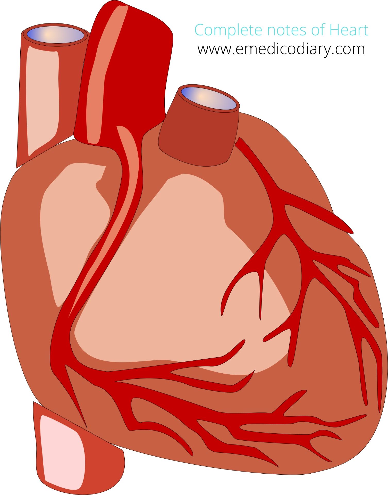
Blood supply
Arterial supply of the heart:
The visceral layer of the heart is supplied with the help of the coronary artery. Which is divided into right and left coronary artery
- Right coronary artery:
The right coronary artery origin from anterior aortic sinus. After arising from ascending aorta it runs forward behind the pulmonary trunk to the right anterior coronary sulcus. Then it reaches up to the junction of the right and inferior border of the heart. Then it turns posteriorly to posterior interventricular groove and terminates by anastomosing with left coronary artery. anatomy of the heart in detail
Branches and distribution
Right conus artery
- It supplies the anterior surface of the pulmonary conus.
Sinuartrial nodal artery
- It supplies to SA node in 60% cases.
- While in 40% cases, the SA node is supplied by the left coronary artery.
Anterior ventricular branches
- They are two or three and supply to the anterior surface of the right ventricle where the marginal branch is largest and supply to the sternocostal surface to apex along the lower margin.
posterior ventricular branches
- They are usually two and supply the diaphragmatic surface of the right ventricle .
Posterior interventricular artery
- It runs in the posterior interventricular groove up to the apex and supplies.
- All the parts of the posterior right ventricle and adjoining part of the posterior left ventricle.
- AV node in 60% cases
- Left coronary artery
The left coronary artery arises from the left posterior aortic sinus of ascending aorta. Then it runs forward and to the left between the pulmonary trunk and left auricle. Then it divides into two artery namely arteries interventricular artery and circumflex artery. Then the anterior interventricular artery runs downward in the anterior intraventricular groove to the apex and from apex it runs posteriorly and terminates by anastomosing with posterior interventricular artery.
Circumflex artery wind around the left margin and continues in the left posterior coronary sulcus up to the posterior IV groove where it terminates by anastomising with the right coronary artery.
Branches and distribution
Anterior interventricular artery:
It supplies
- The greater part of the left ventricle and small adjoining part to the right ventricle
- an anterior part of the interventricular septum.
- A part of left bundle of his.
Circumflex artery:
- Supply to the left margin by left marginal branch up to the apex.
Diagonal artery
- It arises directly from the trunk of the left coronary artery.
Conus artery
- it supplies the pulmonary conus
Arterial Branches
- they supply the left atrium
anatomy of heart in detail
Anastomoses and its importance
It is the point of attachment of two coronary arteries, left and the right coronary artery. It helps in the collateral circulation of the blood if occlusion occurs.
Venous drainage
- Coronary sinus
- Anterior cardiac veins
- Venue cordis minimae
The coronary sinus is the largest vein of the heart lies in the posterior part of the AV groove. It develops from the left horn of sinus venusus. It is 3 cm long which opens into the posterior wall of the right atrium.
Tributaries of the coronary sinus
- Great cardiac
- Middle cardiac vein
- Small cardiac vein
- Posterior vein of the left ventricle
- Posterior vein of the left ventricle
- Oblique vein of the left atrium
- Left marginal vein
- Right marginal vein
Nerve supply
Sympathetic nerve supply
- Derived from upper T3-T5
- Cardio accelerator
- Increase the heart rate
Parasympathetic nerve supply
- Cardio inhibitory
- Slow down the heart rate
- Derived from the vagus nerve
Pain and reflex pathway
- Referred pain= pain feeling at arms and limb during a heart attack
- Radiation pain= radiates to the pathway of nerve
- Shifted pain= pain transfer to next part
Comments (0)