Cranial Nerves (I-VI) (Viva)
6 years ago 3235
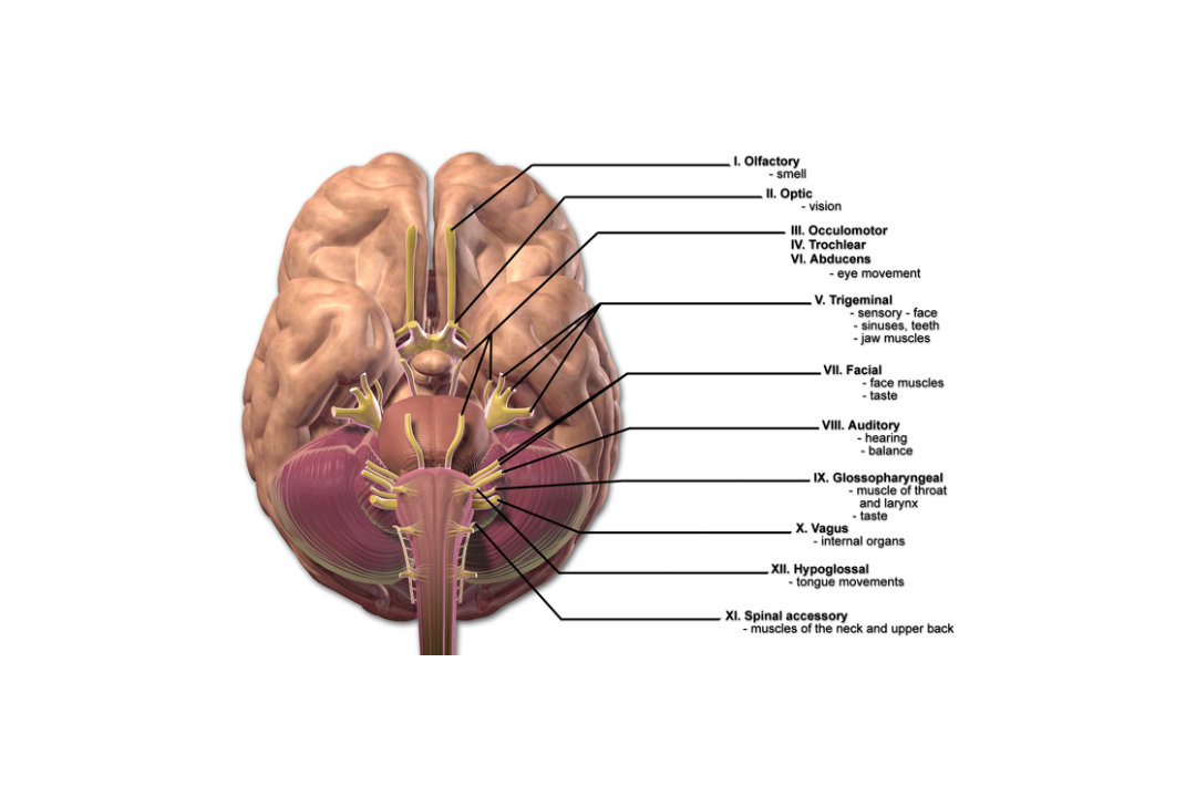
Q.1 Name the cranial nerves.
There are 12 pairs of cranial nerves:
Q.2 How the cranial nerves are classified?
OLFACTORY NERVE
Q.1 Trace the pathway of the olfactory nerve.
Consists of two neurons: Olfactory cells (Receptors)
↓
1st order neuron: Olfactory nerve
↓
Pass through foramina in the cribriform plate of ethmoid
↓
Olfactory bulb
2nd order neuron: Olfactory tract
↓
Divides into
↓
| ↓ | ↓ | ↓ |
| Medial striae ↓ |
Lateral striae ↓ |
Intermediate striae (Sometimes present) ↓ |
| Ends in anterior perforated substance and some fibers cross to the opposite side | anterior perforated substance and primary olfactory cortex | Ends in anterior perforated substance |
| Secondary olfactory cortex |
Q.2 What is the characteristic feature of the olfactory nerve?
The fibers of the olfactory nerve are the central processes of olfactory cells and not the peripheral processes of central ganglion cells.
Q.3 What is hyperosmia?
It is morbid sensitiveness to smell.
Q.4 What is cacosmia?
It is a condition in which a person imagines of non-existent odors.
Q.5 What is the cause of unilateral anosmia (loss of sensation of smell)?
Frontal lobe tumor.
Q.6 What is the cause of bilateral anosmia?
Head injury leading to damage to both olfactory nerves.
OPTIC NERVE
Q.1 What is the length of the optic nerve?
40 mm horizontally and 25 mm vertically is in orbit, 5 mm in the optic canal and 10 mm in the cranial cavity.
Q.2 What are the relations of the intraorbital part of the optic nerve?
Intraorbital part:
Q.3 Trace the optic pathway.
Axons of ganglion cells of retina
↓
Optic nerve
Enters through optic canal
↓
Optic chiasma
(Decussation of fibers occur)
↓
Optic tract
(Has fibers from the nasal half of the macula and retina of the opposite side and temporal half of the same side)
↓
| ↓ Lateral Root | ↓ Medial Root |
| ↓ Terminates in lateral geniculate body | ↓ Terminates in Superior Colliculus, pretectal nucleus, and Hypothalamus |
| ↓ Optic radiation ↓ Pass through the retrolentiform part of the internal capsule ↓ Visual area of cerebral cortex No. 17, 18,19 |
Q.3.1 Trace the pathway of light reflex.
Retina
↓ Optic nerve
↓ Optic chiasma
↓ Optic tract
↓ Lateral geniculate body and pretectal nucleus
↓ Edinger-Westphal nucleus of III cranial nerve
↓ III Cranial nerve
↓ Ciliary ganglion
↓ Short ciliary nerve
↓ Constrictor pupillae muscle
Q.4 What is consensual light reflex?
Constriction of pupil of another eye when the light is flashed on one eye.
Q.5 Why does the consensual light reflex occurs?
Q.6 What is the pathway for accommodation reflex?
Retina
↓ Optic nerve
↓ Optic chiasma
↓ Optic tract
↓ Lateral geniculate body
↓ Optic radiation
↓ Visual area of cortex
↓ Superior longitudinal association tract
↓ Third nerve nucleus
↓ Ciliary ganglion
↓ Ciliaris and sphincter (constrictor) pupillae muscle
Q.7 What are the characteristic features of the optic nerve?
Q.8 How the fibers from the optic tract terminate in the lateral geniculate body?
Fibers from the same eye end in laminae 2,3 and 5 and from the opposite eye end in laminae 1,4 and 6 of the lateral geniculate body. Macular fibers end in the central and posterior parts.
Q.9 What is the Argyll-Robertson pupil?
It is a condition in which pupillary light reflex is absent but the accommodation reflex is present. It is caused by cerebral syphilis. Lesion is in pretectal nuclei.
Q.10 What are the effects of lesions of different parts of the visual pathway?
| Site | Effect |
| Retina | Scotoma (loss of corresponding field) |
| Optic nerve | Blindness of same side Consensual light reflex retained |
| Optic chiasma | |
| -Peripheral lesion: | Binasal hemianopia (Bilateral) |
| -Central lesion | Bitemporal hemianopia. |
| Optic tract, lateral geniculate body, optic radiation | Homonymous hemianopia (Loss of temporal field of one side, and nasal field of other side) No macular sparing |
| Visual cortex | Homonymous hemianopia Macular sparing |
Q.11 Trace the pathway for corneal reflex.
Cornea
↓ Branches of ophthalmic division of V cranial nerve
↓ Main sensory nucleus of V cranial nerve
↓ Secondary fibers to motor nuclei of facial nerve of both sides
↓ Fibers of facial nerve nuclei
↓ Orbicularis oculi muscle
OCULOMOTOR NERVE
Q.1 What are the functional components of the oculomotor nerve?
Q.2 What is the position, subdivisions, and structures supplied by nerves of the oculomotor nucleus?
Position:
At the level of the superior colliculus in the ventromedial part of the central grey matter of midbrain, ventral to the aqueduct. The right and left nuclei fuse to form a midline complex.
Subdivisions:
Q.3 Name the connections of the oculomotor nucleus.
To:
Q.4 What are the relations of the oculomotor nerve in superior orbital fissure?
Nasociliary nerve lies in between and abducent nerve inferolateral to, the two rami of the oculomotor nerve.
Q.5 What is ciliary ganglion and what is its position, connections, and branches?
It is a peripheral ganglion in the course of the oculomotor nerve. Has preganglionic fibers from the Edinger-Westphal nucleus.
Position:
Near apex of orbit between optic nerve and tendon of the lateral rectus muscle.
Connections:
Branches:
Short ciliary nerves 8-10 pierce sclera.
Q.6 What is Weber’s syndrome?
It is a midbrain lesion causing:
Q.7 What are the effects of the intranuclear lesion of the 3rd cranial nerve?
TROCHLEAR NERVE
Q.1 Name the functional components of the IV cranial nerve.
Q.2 What is the position of the trochlear nucleus?
In the ventromedial part of the central grey matter of the midbrain at the level of the inferior colliculus, ventral to the aqueduct. Fibers from the nucleus cross and emerge on the posterior surface of the brainstem just below the inferior colliculus.
Q.3 What is the effect of the lesion of the IV cranial nerve?
Diplopia occurs on looking downwards.
TRIGEMINAL NERVE
Q.1 What are the functional components of the trigeminal nerve?
Q.2 What is the position of the trigeminal nerve nucleus?
It is made up of:
Q.3 What is the position of trigeminal ganglion?
The ganglion is placed in a depression called trigeminal impression in the anterior aspect of the petrous temporal bone and is enclosed in a pouch-like recess of the dura mater.
Q.4 What are the divisions of the trigeminal nerve?
Q.5 What is the distribution of the trigeminal nerve?
Q.6 Name the divisions of ophthalmic nerve and structures supplied by it.
Frontal nerve:
By supratrochlear and supraorbital divisions supply upper eyelid, scalp up to lambdoid suture, and skin of forehead in lower and medial part.
Lacrimal nerve:
To lacrimal gland and lateral part of conjunctiva and skin of the upper eyelid.
Nasociliary nerve:
Eyeball, to ciliary ganglion, medial half of lower eyelid, mucosa and skin of nose and dura of anterior cranial fossa.
Q.7 Name the divisions of the maxillary nerve and its distribution.
Q.8 What is the distribution of the mandibular nerve?
– Nerve to medial pterygoid: Supplies medial pterygoid muscle and gives a branch to the optic ganglion.
– Nerve to tensor palati and tensor tympani.
– Meningeal branch: To dura mater of middle cranial fossa.
– Buccal nerve: Skin of cheek and mucous membrane on its inner aspect.
– Nerve to masseter, temporalis and lateral pterygoid
– Auriculotemporal nerve:
Sensory to skin of temple, auricle, external auditory meatus and tympanic membrane and secretomotor fibers to the parotid gland.
– Lingual nerve:
Mucous membrane of the floor of the mouth and anterior 2/3 of the tongue and secretomotor fibers to the sublingual and submandibular salivary glands.
– Inferior alveolar nerve:
Teeth and lower jaw, the skin over the chin and lower lip and nerve to mylohyoid and anterior belly of digastric.
Q.9 What is the effect of complete unilateral lesion of the trigeminal nerve?
Unilateral anesthesia of face and anterior part of the scalp, auricle and mucous membrane of nose, mouth, and anterior two-thirds of the tongue, with paralysis and wasting of muscles of mastication on the affected side.
Q.10 What is ‘trigeminal neuralgia’?
It is the disease of unknown etiology in which there is sudden severe pain in the area of distribution of the trigeminal nerve.
ABDUCENT NERVE
Q.1 Name the functional components of the abducent nerve.
Q.2 What is the position of VI cranial nerve nucleus?
Upper part of the floor of the fourth ventricle beneath facial colliculus.
Q.3 What is the effect of paralysis of the abducent nerve?
Q.4 What is Raymond syndrome?
It is a pons lesion causing contralateral hemiplegia and paralysis of the abducent nerve on the same side.
Comments (0)