Meninges & CSF (Viva)
6 years ago 5876
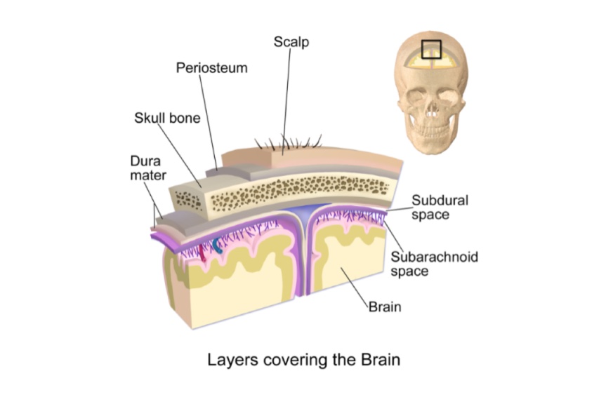
Q.1 What are meninges?
These are the layers of connective tissue covering the brain and spinal cord.
Q.2 What are the layers of the meninges?
The meninges consist of three membranous layers.
The dura mater is also known as ‘Pachymeninges’. The arachnoid and pia mater are together known as ‘Leptomeninges’. The subarachnoid space between the arachnoid and pia mater contains cerebrospinal fluid.
Q.3 What is the developmental origin of meninges?
Leptomeninges: From neural crest.
Pachymeninges: From mesoderm surrounding neural tube.
Q.4 What are the layers of the dura mater?
Dura mater is the thickest and toughest membrane covering the brain and consists of two layers:
Q.5 To what structures the endosteal layer is attached.
It is attached to:
Q.6 What are the structures covered by the endosteal layer other than the brain?
It provides tubular sheaths for cranial nerves and fuses with epineurium except the optic nerve, where sheaths are derived from meninges.
Q.7 What are meningocytes and what is their function?
Meningocytes are mesothelial cells found in:
Functions:
Q.8 Name the coverings of the spinal cord.
Q.9 What are the folds of dura mater?
These are formed by the meningeal layer of the dura mater around the brain. These projects inwards and divide the cranial cavity into different compartments.
These are:
Q.10 What is the ‘falx cerebri’?
It is a fold of dura mater, which is sickle-shaped and occupies the median longitudinal fissure between two cerebral hemispheres.
Q.11 What are the venous sinuses enclosed by the falx cerebri?
Q.12 What is ‘tentorium cerebelli’?
It is a tent-shaped fold of dura mater, forming the roof of the posterior cranial fossa. It separates the cerebellum from the occipital lobes of the cerebrum. It lies at right angles to falx cerebri.
Q.13 What are sinuses enclosed by the tentorium cerebelli?
The attached margin encloses the transverse sinus in the posterior part and superior petrosal sinus in the anterolateral part.
Q.14 Where the straight sinus is situated?
At the junction of the lower edge of falx cerebri with the upper surface of the tentorium cerebelli. It lies between the left and right layers of falx cerebri which become continuous with corresponding half of the upper layer of the tentorium cerebelli. The lower layer of tentorium cerebelli passes transversely across the midline without interruption.
Q.15 How the straight sinus is formed and terminates?
Anteriorly it receives inferior sagittal sinus and great cerebral vein and posteriorly it terminates by becoming continuous with the transverse sinus of the side opposite to that with which the superior sagittal sinus is continuous, usually left side.
Q.16 What is ‘trigeminal cave’?
Trigeminal or Meckel’s cave is a recess of dura mater, formed by the inferior layer of tentorium cerebelli, over the trigeminal ganglion, on the anterior surface of the petrous temporal bone.
Q.17 What is ‘flax cerebelli’?
It is a small sickle-shaped fold in sagittal plane projecting forwards into the posterior cerebellar notch.
Q.18 Which sinus is enclosed by falx cerebelli?
Occipital sinus lies along the posteriorly attached part.
Q.19 What is ‘diaphragma sellae’?
It is a circular horizontal fold of the dura mater forming the roof of the hypophyseal fossa, in the middle cranial fossa.
Q.20 What is the structure transmitted by the central aperture of diaphragm sellae?
Pituitary stalk (Infundibulum)
Q.21 What are the characteristics of venous sinuses of the skull?
Q.22 What are different venous sinuses of the skull?
The venous sinuses of the skull can be divided into two broad groups:
| Paired: | Unpaired: |
|
– Cavernous – Superior petrosal – Inferior petrosal – Transverse – Sigmoid – Sphenoparietal – Petrosquamous and – Middle meningeal sinus |
– Superior sagittal – Inferior sagittal – Straight – Occipital – Anterior intercavernous – Posterior intercavernous – Basilar plexus veins. |
Q.23 Where is the cavernous sinus is situated?
It is situated in the middle cranial fossa on either side of the body of the sphenoid bone.
Q.24 What are the relations of cavernous sinus?
The relations can be divided into 3 broad subdivisions :
– Oculomotor nerve
– Trochlear nerve
– Ophthalmic nerve
– Maxillary nerve
– Trigeminal ganglion.
Superiorly:
- Optic tract
- Internal carotid artery
- Anterior perforated substance.
Inferiorly:
- Foramen lacerum
- Junction of body and greater wing of sphenoid.
Medially:
- Hypophysis (Pituitary gland)
- Sphenoidal air sinus.
Laterally:
- Temporal lobe with uncus.
Anteriorly:
- Superior orbital fissure
- Apex of orbit.
Posteriorly:
- Apex of petrous temporal
- Crus cerebri of the midbrain.
- Internal carotid artery
- Abducent nerve
Q.25 Name the communications of cavernous sinus.
These are:
Q.26 What are the tributaries of cavernous sinus?
– Sphenoparietal sinus
– Frontal trunk of middle meningeal vein.
– Superior middle cerebral vein
– Inferior cerebral veins.
– Superior ophthalmic vein.
– Inferior ophthalmic vein.
– Central vein of retina
Q.27 What is the commonest cause of thrombosis of cavernous sinus?
Infection of the danger area of the face, nasal cavities, and paranasal air sinuses.
Q.28 What do you understand by the term ‘confluence of sinuses’?
This is the posterior dilated end of the superior sagittal sinus lying on the right side of the internal occipital protuberance. It continues as corresponding transverse sinus and it is connected to the opposite transverse sinus and straight sinus and drains the occipital sinus.
Q.29 What is the characteristic feature of pia mater?
It is a highly vascular layer and is closely adherent to the brain extending into the sulci, but the arachnoid mater does not do so and jumps across the sulci. So the subarachnoid space extends into the sulci.
Q.30 What are arachnoid villi?
These are the finger-like processes of the arachnoid tissue which project into cranial venous sinuses. Their function is to absorb CSF into the bloodstream.
Q.31 What are Pacchionian bodies?
Also called arachnoid granulations. These are aggregations of arachnoid villi clumped together. Found in adults. They are most numerous in relation to superior sagittal sinus.
Q.32 What is ‘Tela choroidea’ and ‘choroid plexuses’? What are their importance?
The folds of highly vascular pia mater projecting into ventricles are tela choroidea.
Cavity of the ventricle is lined by ependyma. The masses of pia mater covered by ependyma are referred to as choroid plexuses.
Importance: At these sites, the CSF is secreted into the ventricles of the brain.
Q.33 What are ‘Cisterns’? What are their functions?
These are communicating pools formed by the subarachnoid space at the base of the brain and around the brainstem.
Function: These reinforce the protective effect on the vital centers in the medulla.
Q.34 What are the communications of subarachnoid space?
It communicates with the ventricular system of the brain by:
Q.35 What is CSF?
It is a clear fluid found in the subarachnoid space of the brain and spinal cord, a ventricular system of the brain and the central canal of the spinal cord.
Q.36 Where is CSF formed?
It is formed by choroid plexuses of the ventricles of the brain by an active secretory process.
Q.37 What is the pathway of the circulation of CSF?
| Lateral ventricles ↓ Foramina of Monro Third ventricle ↓ Cerebral aqueduct ↓ Fourth ventricle Foramina of Magendie and Foramina of Luschka. ↓ Subarachnoid space around the brain and spinal cord ↓ Absorbed by arachnoid villi, perineural lymphatics around cranial nerve |
Q.38 How a sample of CSF obtained?
Q.39 What are the functions of CSF?
It is protective and nutritive to the CNS.
Q.40 What is hydrocephalus?
It is the dilatation of the ventricular system and enlargement of head due to obstruction of flow of CSF within ventricular system in children.
Q.41 What is Queckenstedt’s test?
Done to detect whether there is a blockade to the circulation of CSF in subarachnoid space of spinal cord.
Anatomical basis of test:
Any increase in intracranial pressure raises the pressure of CSF. This increase is transmitted to CSF in spinal subarachnoid space.
Compression of both internal jugular veins above the sternal ends of clavicles dams back blood in the skull and so raises the intracranial pressure. Should a part of the spinal subarachnoid space be completely cut off from above by a tumor, this increase of pressure will not be transmitted to the part of the subarachnoid space below the tumor.
Q.42 What is the commonest cause of extradural hemorrhage?
Rupture of the anterior division of the middle meningeal artery.
Q.43 What is the commonest cause of subdural hemorrhage?
Rupture of superior cerebral vein at its entry into superior sagittal sinus.
Comments (0)