Forebrain (Viva)
6 years ago 4249
Q.1 What are the different parts of the brain?
The brain is divided into three parts:
Q.2 How the brain develops?
The brain develops from the cranial part of the neural tube. The cavity of the developing brain shows three dilatations.
Craniocaudally, these are prosencephalon, mesencephalon, and rhombencephalon.
FOREBRAIN
Q.1 What are the subdivisions of the forebrain?
TELENCEPHALON
Q.1 What are the different lobes of the cerebrum?
This division is done by:
Three sulci: Central, lateral, occipitoparietal.
Two imaginary lines:
One from the parietooccipital sulcus to the preoccipital notch and the second is backward continuation of the posterior ramus of the lateral sulcus before it turns upwards and meets the first line.
Q.2 What are the structural divisions of the cerebral cortex?
Q.3 What are the functional divisions of the cerebral cortex?
The cortex is divided into motor and sensory areas.
Q.4 What is the motor area of the cerebral cortex?
In precentral gyrus on the superolateral surface and in the anterior part of paracentral lobule on the medial surface of the cerebral hemisphere. It corresponds to areas 4 of Brodmann.
Q.5 How the lateral and anterior corticospinal tracts are formed?
The corticospinal fibers from the cerebral cortex descend and at the lower end of the medulla 80% cross to the opposite side forming the lateral tract. Fibers that do not cross from the anterior corticospinal tract and at appropriate levels of spinal cord cross to the opposite side. So both tracts ultimately connect the cerebral cortex of one side with the opposite half of the spinal cord ending in ventral grey column neurons.
Q.6 How the body parts are represented in the cerebral cortex?
Q.7 What is Broca’s area?
It is motor speech area No. 44, 45 which controls the speech. It lies in the inferior frontal gyrus. Lesion of this area produces ‘motor aphasia’.
Q.8.0 What is the effect of lesion at area 6,8 of the Frontal lobe?
Loss of horizontal conjugate movements of the eyes.
Q.8 What is the ‘sensory area’?
It is located in the postcentral gyrus and corresponds to areas 1, 2, and 3 of Brodmann.
Q.9 What are the boundaries of interpedicular fossa?
This area lies anterior to the midbrain.
Boundaries:
In front: Optic chiasma
On sides: Optic tracts
Q.10 What are the boundaries of anterior perforated substance?
It is a triangular area lying on each side of the optic chiasma.
Boundaries:
Anterolateral: Lateral olfactory stria
Posterolateral: Uncus
Q.11 Name the constituents of the limbic system.
Q.12 What are the functions of the limbic system?
It controls:
Q.13 Where the hippocampus is situated?
The hippocampus forms a longitudinal projection occupying the greater part of the floor of the inferior horn of the lateral ventricle. It is the superior limb of the ‘S’ of the cerebral cortex that lies between the choroid fissure above and the hippocampal fissure below.
Q.14 What are the communications of the lateral ventricle?
Each lateral ventricle communicates with the third ventricle through an interventricular foramen or foramen of Monro.
Q.15 What are the different parts of the lateral ventricle?
Each lateral ventricle is made up of:
Q.16 What are the constituents of the white matter of the cerebrum?
It consists of myelinated fibers that connect various parts of the cortex and other parts of the CNS.
Q.17 What are the different types of fibers of white matter?
Three types:
Q.18 What are the different commissures of the cerebrum?
Q.19 What are the different parts of the corpus callosum?
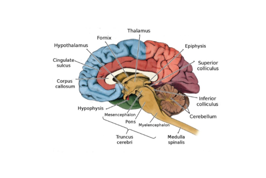
DIENCEPHALON
Q.1 Name the parts of the diencephalon.
Q.2 Name the cavity of diencephalon.
Third ventricle
Q.3 What are the different parts and nuclei of the thalamus?
White matter:
Grey matter:
Q.4 What are the afferents and efferents to the thalamus?
Afferents:
Efferents:
Q.5 What are the functions of the thalamus?
Q.6 What is the characteristic feature of the structure of the lateral geniculate body?
It is six-layered. Layers 1,4 and 6 receive contralateral optic fibers and layers 2,3 and 5 ipsilateral fibers.
Q.7 What is the pineal body?
It is a small conical body projecting downwards in the posterior wall of the third ventricle, just above the superior colliculi of the midbrain.
Q.8 What is ‘brain sand’?
Calcareous concretions appear in the pineal body after 17 years of life and form aggregations. These are called brain sand or corpora arenacea. They appear as radioopaque structures in X-ray.
Q.9 What is the clinical importance of Pineal concretions?
Normally the pineal concretions appear as midline structure in X-ray. They are shifted in cases of head injury.
Q.10 What is the function of the pineal body?
Q.11 Which cranial nerve is likely to be paralyzed in tumors of the pineal body?
It presses on the tectum of the midbrain damaging the oculomotor nucleus and leading to paralysis of the oculomotor nerve.
Q.12 Why hypothalamus is called the ‘head ganglion of the autonomic nervous system’?
Because it controls the various autonomic activities of the body. Sympathetic by caudal part and parasympathetic by cranial part.
Q.13 What is the position of the hypothalamus?
It is present at the base of the brain and forms the floor and lateral wall of the third ventricle.
It is related to:
Anteriorly: Lamina terminalis
Posteriorly: Subthalamus and Tegmentum of midbrain.
Laterally: Internal capsule and Subthalamus.
Q.14 Which structures are related to the hypothalamus in the floor of the third ventricle?
Q.15 How does the preoptic region differs from the rest of the hypothalamus?
Preoptic region is a derivative of the telencephalon.
Q.16 What is the function of habenular nuclei?
These are regarded as cell stations in the olfactory and visceral pathways.
Q.17 What are the functions of the hypothalamus?
• Endocrine control:
By releasing or release inhibiting hormones, it regulates the functions of various endocrine glands of the body.
• Neurosecretion:
Oxytocin and ADH are secreted by the hypothalamo-hypophyseal tract to the posterior pituitary.
• Control of sexual behavior and reproduction through anterior pituitary.
• Regulation of food and water intake:
Lateral zone is responsible for hunger, thirst, and drinking, and the medial zone for satiety.
• Temperature regulation
• Control of emotional behavior.
• Maintains circadian rhythm of body: By suprachiasmatic nucleus.
• Control of autonomic functions.
Q.18 What are the recesses of the third ventricle?
These are the extensions of the cavity of the third ventricle.
These are:
Q.19 Name the structures forming the lateral wall of the third ventricle.
From above downwards
Q.20 Name the structures present on the floor of the third ventricle.
INTERNAL CAPSULE
Q.1 What are the different parts of the internal capsule?
Q.2 What is the arrangement of corticospinal fibers in the posterior limb of the internal capsule?
The arrangement of fibers from anterior to posterior is upper limb, trunk, and then lower limb.
Q.3 Where is the clinical importance of the blood supply of the internal capsule?
Lateral striate artery (Charcot’s artery) supplying the internal capsule is the commonest site of hemorrhage in cases of hypertension and it leads to the paralysis of the opposite half of the body (hemiplegia), depending on which side is involved in hemorrhage.
BASAL GANGLIA
Q.1 What are basal ganglia?
These are masses of grey matter situated in cerebral hemispheres forming part of the extrapyramidal system.
These are:
Caudate nucleus and lentiform nucleus together constitute corpus striatum.
Q.2 What are the morphological divisions of corpus striatum?
The putamen and caudate nucleus form neostriatum, globus pallidus forms paleostriatum and amygdaloid body forms archistriatum.
Q.3 What is the function of the corpus striatum?
It is an important integrating center in motor activity.
Comments (0)