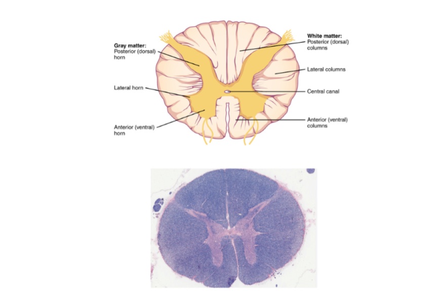
CNS-Spinal Cord (Viva)
Q.1 What are the divisions of the nervous system?
Anatomically the nervous system is made up of:
- Central nervous system (CNS): Consisting of the
– Brain and
– Spinal cord
- Peripheral nervous system (PNS): Consisting of
- Somatic (Cerebrospinal) nervous system
- Autonomic (Splanchnic) nervous system
Q.2 What are the constituents of the somatic nervous system?
It consists of 12 pairs of cranial nerves and 31 pairs of spinal nerves.
Q.3 What are the functions of the somatic nervous system?
It is concerned with the response of the body to the external environment.
Q.4 Name the constituents of the ‘autonomic nervous system’?
It consists of the sympathetic and parasympathetic nervous system.
Q.5 What are the functions of the autonomic nervous system?
It is mainly concerned with the control of the internal environment of the body, e.g. regulation of heart, bronchial tree, gut, and glands of the alimentary tract.
Q.6 What is the main difference between the somatic and autonomic nervous systems?
The efferent fibers of the somatic nervous system reach the effectors without interruption while the efferent fibers of ANS first relays in a ganglion and then postganglionic fibers pass to the effectors.
SPINAL CORD
Q.7 What is the extent of the spinal cord?
It extends from the upper border of the atlas vertebra to the lower border of L1.
Q.8 How the spinal cord develops?
It develops from the caudal tubular part of the neural tube, which gradually increases in length.
Q.9 What are the age changes in the length of the spinal cord?
Up to 3rd month of fetal life: Spinal cord occupies the full extent of the vertebral canal.
At birth: At the level of L3 vertebra.
At adolescence: At the level of the intervertebral disc between L1 and L2 vertebra.
Q.10 Name the arteries supplying the spinal cord?
• Anterior spinal artery:
One, in anterior median fissure
• Posterior spinal artery:
Two, along the posterolateral sulcus, i.e. along the line of attachment of dorsal nerve roots.
• Arterial vasocorona:
Arterial plexus in pia mater covering the spinal cord.
• Radicular arteries:
Reach cord along roots of spinal nerves.
Q.11 Which artery supplies the greater part of the cross-section of the spinal cord?
Anterior spinal artery
Q.12 What is the venous drainage of the spinal cord?
The veins draining the spinal cord are arranged in six longitudinal channels.
Anteromedian and posteromedian lying in the midline and anterolateral and posterolateral that are paired. These are interconnected by a plexus, venous vasocorona, these drain into radicular veins which in turn drain into epidural venous plexus and which drains into the external vertebral venous plexus through intervertebral and basivertebral veins.
Q.13 What are ‘arteries of Adamkiewicz’?
These are the anastomotic arteries between anterior and posterior spinal arteries at the level of T1 and T11.
Q.14 Name the ‘descending’ and ‘ascending’ tracts of the spinal cord?
Descending tracts: Motor in function:
- Lateral corticospinal
- Anterior corticospinal
- Rubrospinal
- Vestibulospinal
- Olivospinal
- Tectospinal
- Medial reticulospinal
- Lateral reticulospinal.
Ascending tracts: Sensory in functions:
- Fasciculus gracilis
- Fasciculus cuneatus
- Posterior spinocerebellar
- Anterior spinocerebellar
- Lateral spinothalamic
- Anterior spinothalamic
- Spinotectal
- Spino-olivary
Q.15 What is the function of interneurons?
- Axon of an interneuron may form a number of branches, which synapse with a number of efferent neurons. So, an impulse in a single afferent neuron may result in an effector response by a number of efferent neurons.
- Afferent impulses from different afferent neurons may converge on a single afferent neurons through interneurons. These impulses may be facilitatory or inhibitory.
- Afferent neurons may form contact with efferent neurons in the opposite half of the spinal cord or in the higher or lower segments of the cord through the interneuron.
Q.16 Trace the pathway of the posterior column ascending tract.
Receptors: Sensory end organs in various tissues.
Peripheral process of dorsal root neurons form the afferent fibers of peripheral nerves.
First-order neuron:
Central processes of neurons in dorsal nerve root ganglia. The fibers ascend in the spinal cord as posterior column tracts, up to the lower part of the medulla and end in nucleus gracilis and nucleus cuneatus.
Second-order neuron:
Neurons in nucleus gracilis and nucleus cuneatus. The axons cross the midline (sensory decussation) and run upwards as medial lemniscus to end in the thalamus, passing through the medulla, pons, and midbrain.
Third-order neuron:
Neurons in thalamus. Gives axons to the somatosensory area of the cerebral cortex passing through the internal capsule and corona radiata.
Q.17 What are the tracts of the posterior column?
- Fasciculus gracilis and
- Fasciculus cuneatus.
Q.18 What are the sensations carried by the posterior column?
- Deep touch and pressure
- Tactile localization
- Tactile discrimination
- Stereognosis
- Sense of vibration
- Sense of position and movements of different parts of the body (Proprioceptive impulses).
Q.19 What are the sensations carried by spinothalamic tracts?
- Anterior spinothalamic tract: Sensation of crude touch and pressure.
- Lateral spinothalamic tract: Sensation of pain and temperature.
Q.20 What are the functions of spinocerebellar tracts?
These carry proprioceptive impulses arising in muscle spindles, Golgi tendon organs, and other proprioceptive receptors of lower limbs.
Dorsal tract: Impulses concerned with fine coordination of muscles controlling posture and movements of individual muscles.
Ventral tract: Concerned with movements of limb as a whole.
Q.21 What is the function of various descending spinal tracts?
These influence the activity of ventral column neurons both alpha and gamma, through internuncial neurons, affecting both contraction and tone of skeletal muscle. They also influence the transmission of afferent impulses through ascending tracts.
Q.22 What are `ligamentum denticulata'? What is their function?
These are toothed processes extending from pia to dura, pushing the arachnoid before them. They leave the pia midway between anterior and posterior nerve roots and serve to suspend the spinal cord in the midline.
Q.23 What is ‘conus medullaris’?
It is the lower end of the spinal cord which is conical. The apex of the conus continues downwards as filum terminale, up to the first coccygeal space.
Q.24 What is ‘cauda equina’?
The spinal cord gives rise to spinal nerves which pass out through intervertebral foramina. Below the L1 vertebra, nerve roots become more and more oblique to reach respective intervertebral foramina. The bundle of lumbar and sacral nerve roots below the termination of the spinal cord is termed cauda equina.
Q.25 What is cauda equina syndrome?
Compression of cauda equina gives rise to flaccid paraplegia, saddle anesthesia, which is known as cauda equina syndrome.
Q.26 What are the effects of the complete transection of the spinal cord?
In the region below section, there is a complete loss of sensation with flaccid muscle paralysis.
Q.27 What is Tabes dorsalis?
It is a degenerative disease of posterior columns and posterior nerve roots, which is characterized by loss of proprioception (position sense).
Q.28 What is Brown-Séquard’s syndrome?
It occurs in the hemisection of the spinal cord.
It is characterized by:
- Paralysis of the affected side below the lesion (Corticospinal tract).
- Loss of proprioception and fine discrimination on affected side below lesion (Fasciculus cuneatus and gracilis).
- Loss of pain and temperature sense on the opposite side below the lesion (Spinothalamic tract).
Q.29 At what site lumbar puncture is done?
Lumbar puncture is done to withdraw CSF from subarachnoid space at the level between L3 and L4 vertebra.

Comments (0)