Joints of Head & Neck (Viva)
6 years ago 3654
Q.1 What type of joint is a temporomandibular joint (TM joint)?
Condylar variety of synovial joint
Q.2 Name the ligaments of the T-M joint.
Q.3 What are the characteristic features of articular disc?
Q.4 What is the developmental origin of sphenomandibular ligament?
It is a remnant of the dorsal (cephalic) end of Meckel’s cartilage.
Q.5 Name the structures piercing sphenomandibular ligament.
Mylohyoid nerve and vessels
Q.6 What is the nerve supply of the TM joint?
Q.7 Name the muscles of T-M joint (Muscles of mastication).
Q.8 What is the developmental origin of muscles of mastication?
Mesoderm of first branchial arch
Q.9 What are the structures passing between two heads of lateral pterygoid?
Q.10 What are the movements of TM joints?
Q.11 Describe movements which occur in chewing.
Head of one side of the mandible with articular disc
Q.12 What are the different muscles producing movements of the jaw joint?
Depression:
Lateral pterygoid of both sides with digastric, geniohyoid and mylohyoid
Elevation:
Masseter, temporalis, and medial pterygoid of both sides
Protraction:
Lateral and medial pterygoids with masseter
Retraction:
Posterior fibers of temporalis with digastric and geniohyoid
Chewing:
Medial and lateral pterygoids of each side acting alternately.
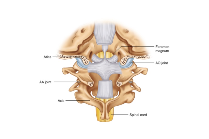
ATLANTO-OCCIPITAL JOINT
Q.1 What is the variety of Atlantooccipital joint?
Ellipsoid variety of synovial joint
Q.2 What are the articular surfaces of the Atlanto-occipital joint?
From above: Occipital condyles.
From below: Superior articular facets of the atlas vertebra.
Q.3 What are the movements, possible at this joint?
ATLANTO-AXIAL JOINT
Q.1 What type of this joint is?
Q.2 Name the ligaments connecting atlas, axis, and occipital bones.
Q.3 Name the ligaments connecting the dens of the axis with the occipital bone.
Q.4 Which movement takes place at atlanto-axial joint?
Side to side movement
Q.5 How the death in hanging occurs?
Due to dislocation of dens of axis following rupture of the transverse ligament of the atlas, which then crushes the spinal cord and medulla, i.e. vital centers
Comments (0)