Mandible & Hyoid Bone (Viva)
6 years ago 4476
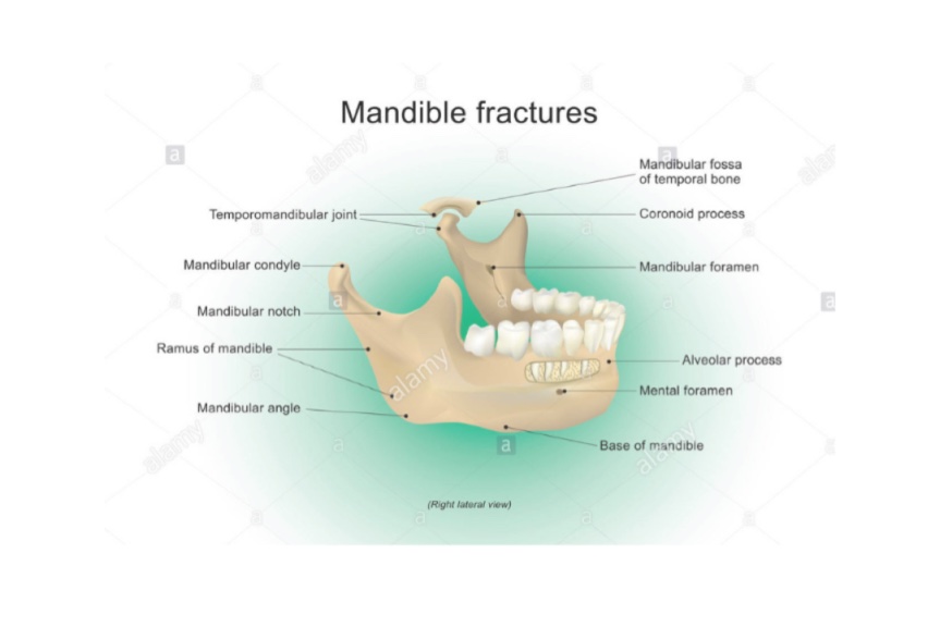
Q.1 Name the structures transmitted by mental foramen.
Mental nerve and vessels
Q.2 Name the structures present in the submandibular fossa.
Q.3 What structures are attached to genial tubercles?
Q.4 Name the structures attached to the angle of the mandible.
Q.5 Name the structures related to the neck of the mandible.
On lateral aspect:
Parotid gland below the attachment of the lateral ligament of the jaw joint.
On medial aspect:
Q.6 What are the changes in the position of mental foramen with age?
The mental foramen at birth opens below sockets for deciduous molar teeth near the lower border. The foramen gradually shifts upwards and in adults, it opens midway between upper and lower borders.
In old age, due to the absorption of the alveolar border, the mental foramen lies close to the alveolar border.
Q.7 What is the characteristic feature of the ossification of the mandible?
It ossifies partly in membrane and partly in cartilage.
Parts ossifying in membrane:
Body of mandible except incisive part and lower half of ramus up to mandibular foramen.
Parts ossifying in cartilage:
Incisive part below incisor teeth, coronoid and condyloid processes, and upper half of ramus above mandibular foramen.
Q.8 How will you determine the sex to which the mandible belongs?
| Features | Male | Female |
| General size | Larger and thicker | Smaller and thinner |
| Height of body | Greater | Lesser |
| Angle of mandible | Lesser, Everted | Greater, Inverted |
| Chin | Quadrilateral | Rounded |
| Inferior border of body of mandible |
Irregular | Smooth curve |
| Condyles | Larger | Smaller |
Q.9 What is the commonest site of fracture of the body of mandible?
At the level of canine socket
HYOID BONE
Q.1 What is the level of hyoid bone?
C3 vertebra behind and base of mandible in front.
Q.2 Name the structures attached to the anterior surface of the body of the hyoid.
Insertion to: Geniohyoid and mylohyoid.
Origin to: Hyoglossus.
Below mylohyoid: Investing fascia.
Q.3 Name the structures attached to the lower border of the hyoid body?
Sternohyoid: Medial
Omohyoid, superior belly: Lateral
Thyrohyoid: Below omohyoid
Pretracheal fascia.
Q.4 What structures are attached to greater cornua of hyoid?
Upper surface:
Medial border:
Lateral border:
Q.5 What structures are attached to lesser cornua of hyoid?
Q.6 What is the developmental origin of hyoid bone?
Upper half of body and lesser cornua: Cartilage of second pharyngeal arch
Lower half of body and greater cornua: Cartilage of third pharyngeal arch.
Comments (0)