Skull Bones (Viva)
6 years ago 7052
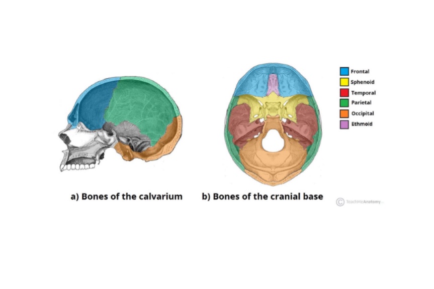
Q.1 What is the total number of bones in the skull?
Twenty-two
Q.2 How the skull is held in a normal anatomical position?
By considering any one of the following two planes:
Q.3 What are sutures?
The joints between the various skull bones. These are immovable and fibrous in type.
Q.4 What are the different sutures seen in the superior view (Norma verticalis) of the skull.
Q.5 What is vertex?
It is the highest point on the sagittal suture.
Q.6 What is Bregma?
Meeting point between coronal and sagittal sutures. In foetal skull, represents anterior fontanelle, which closes at the age of 1½ years.
Q.7 What is Lambda?
Meeting point between sagittal and lambdoid sutures. In foetal skull, represent the posterior fontanelle, which closes by the age of 2-3 months.
Q.8 Name the structures attached to external occipital protuberances?
Trapezius, in the upper part.
Ligamentum nuchae, in the lower part.
Q.9 What is ‘Occipital point’?
Median point just above inion (most prominent part of external occipital protuberance) which is farthest from the glabella.
Q.10 What is ‘glabella’?
The meeting point of two superciliary arch in midline.
Q.11 What is ‘Nasion’?
Median point at the root of the nose, where the internasal suture meets with the frontonasal suture.
Q.12 Name the bones forming medial orbital margin.
Frontal process of maxilla, mainly.
Nasal part of the frontal bone, in the upper part.
Q.13 Name the sutures present in Norma frontalis?
Q.14 What is ‘Jugal point’?
Anterior end of the upper border of the zygomatic arch.
Q.15 What are the boundaries of the supramental triangle?
Above: Supramastoid crest
Front: Posterosuperior margin of the external auditory meatus.
Behind: Vertical tangent to posterior margin of meatus.
Q.16 What is the significance of tegmen tympani?
It is a thin plate of bone formed by petrous temporal bone. It separates the middle cranial fossa from the cavities of the middle ear, auditory tube, and mastoid antrum.
Q.17 Name the structures attached to the mastoid process.
From before backward:
Q.18 What are the relations of the styloid process?
Laterally: Parotid gland.
Medially: Internal jugular vein.
Base: Ensheathed by tympanic plate. Related to facial nerve.
Apex: Posterior border of ramus of mandible, laterally
Q.19 Name the structures attached to the styloid process.
Muscle:
Ligaments:
Q.20 What is ‘Pterion’?
H-shaped suture seen in norma lateralis formed by the frontal, parietal, sphenoid, and temporal bone.
Q.21 What is ‘asterion’?
The point at which parietomastoid suture (formed by mastoid part of the temporal bone with parietal bone) and occipitomastoid suture (formed by mastoid part of the temporal bone with occipital bone) meet.
Q.22 Name the structures related to pterion.
Q.23 What are the boundaries of temporal fossa?
Superior: Temporal line
Inferior: Zygomatic arch
Floor: By parts of the frontal, parietal, squamous temporal, and greater wing of sphenoid bone.
Anterior wall: Mainly by the temporal surface of zygomatic bone. Also by greater wing of sphenoid and frontal bone.
Q.24 What is the origin, insertion, and nerve supply of temporalis muscle?
Origin: Floor of temporal fossa
Insertion: Coronoid process of mandible
Nerve supply: Deep temporal nerve, branch of the anterior division of the mandibular nerve.
Q.25 What are the boundaries of infratemporal fossa?
Roof: Mainly by greater wing of sphenoid. Also squamous temporal bone.
Medial: Pterygoid process of sphenoid
Anterior: Posterior surface of maxilla.
Q.26 What are the communications of infratemporal fossa?
Q.27 What are the contents of ‘infratemporal fossa’?
Q.28 Name the bones forming hard palate.
Anterior 2/3: Palatine processes of maxilla.
Posterior 1/3: Horizontal plates of palatine bones.
Q.29 Name the structures attached to the lateral pterygoid plate.
Lateral surface: Lower head of lateral pterygoid.
Medial surface: Deep head of medial pterygoid.
Posterior border: Pterygospinous ligament.
Q.30 Why the upper surface of the body of sphenoid is called ‘Sella turcica’?
Because it is hollowed out in the form of a ‘Turkish saddle’.
Q.31 What lodges the hypophysis (Pituitary gland)?
Hypophyseal fossa of sphenoid bone.
Q.32 What are the boundaries of ‘superior orbital fissure’?
Above and medially: Lesser wing of sphenoid.
Below and laterally: Greater wing of sphenoid.
Q.33 What are the boundaries of inferior orbital fissure?
Above and laterally: Greater wing of sphenoid.
Below and medially: Orbital surface of the maxilla.
Q.34 What are the relations of the ‘spine of sphenoid’?
Lateral: Auriculotemporal nerve.
Medial: Chorda tympani nerve. Auditory tube.
Tip: Attachment to sphenomandibular ligament.
Anterior: Origin of fibers of tensor palati.
Q.35 Name the structures passing through internal acoustic meatus.
Q.36 What are the boundaries of the optic canal?
Lateral: Lesser wing of sphenoid
Medial: Body of sphenoid.
Q.37 What structures are transmitted through the optic canal?
Q.38 Name the structures passing through superior orbital fissure?
It is divided into three parts by a common tendinous ring of Zinn.
It transmits:
– Lacrimal nerve, Branch of ophthalmic division
– Frontal nerve, of the trigeminal nerve
– Trochlear nerve
– Recurrent branch of the ophthalmic artery and
– Superior ophthalmic vein.
– Upper and lower division of oculomotor nerve,
– Nasociliary nerve, branch of the ophthalmic division of V nerve and
– Abducent nerve.
– Inferior ophthalmic vein.
Q.39 What structure passes through Foramen rotundum?
Maxillary nerve.
Q.39.1 What structures pass through the Foramen ovale?
Q.40 Name the structures passing through Foramen spinosum.
Q.41.0 What are the boundaries of Foramen magnum?
Anterior: Basiocciput
Posterior: Squamous occipital
On each side: Condylar part of occipital bone
Q.41 Name the structures passing through Foramen magnum.
– Lower part of medulla.
– Tonsils of cerebellum.
– Meninges.
– Apical ligament of dens.
– Membrana tectoria.
– Superior band of cruciform ligament.
– Spinal accessory nerve.
– Vertebral arteries
– Sympathetic plexus.
– Posterior spinal arteries.
– Anterior spinal arteries.
Q.42 Name the structure transmitted by mastoid foramen.
Emissary vein connecting sigmoid sinus to occipital vein.
Q.43 Name the structure transmitted by sphenopalatine foramen.
Nasopalatine nerve and vessels.
Q.44 What are the contents of the Hypoglossal canal?
Q.45 Name the structures passing through Jugular foramen?
– Inferior petrosal sinus
– IX, X, and XI cranial nerves.
– Meningeal branch of ascending pharyngeal artery.
– Lower end of sigmoid sinus
– Meningeal branch of the occipital artery.
– Emissary vein connecting sigmoid sinus to occipital vein.
Q.46 Name the structures transmitted by the carotid canal.
Q.47 Name the structures transmitted by inferior orbital fissure.
Q.48 Deep petrosal and greater petrosal nerve passes through which foramen of the skull?
Foramen lacerum
Q.49 Name the structures passing through stylomastoid foramen?
Q.50 What are Wormian bones?
These are small irregular bones found in the region of fontanelles and are formed by the additional ossification centers.
Q.51 Which skull bone is formed by intramembranous ossification?
Frontal, parietal, zygomatic, palatine, nasal, lacrimal, maxilla and vomer.
Q.52 Which skull bone is formed partly in cartilage and partly in membrane?
Occipital, sphenoid, temporal, and mandible.
Q.53 Which skull bone is formed entirely in cartilage?
Ethmoid and inferior nasal concha.
Q.54 What is the Cephalic index?
It is the ratio of the breadth (widest diameter) and length (longest diameter) of the skull.
Q.55 What is the clinical importance of the Cephalic index?
Q.56 What is ‘Scaphocephaly’?
Boat-shaped skull resulting from early union of the sagittal suture.
Q.57 What is ‘Acrocephaly’?
Pointed skull as a result of the early union of the coronal suture.
Comments (0)