Ear (Viva)
6 years ago 3141
Q.1 What are the parts of the external ear?
Q.2 What is the nerve supply of auricle?
Lateral surface:
– Anterosuperior part including tragus:
Auriculotemporal (Branch of mandibular division of Trigeminal nerve)
– Posteroinferior part including lobule:
Greater auricular (C2,3)
Cranial Surface
– Upper 1/3: Lesser occipital (C2)
– Lower 2/3: Greater auricular (C2,3)
– Eminentia conchae: Auriculotemporal nerve
Concavity of conchae on external surface: Auricular branch of vagus.
To auricular muscles: Facial nerve.
Q.3 What is the shape of external acoustic meatus?
It follows an S-shaped course. Cartilaginous part, first passes medially, forwards and upwards. It then passes medially, backward, and upwards. Bony part runs medially, forwards and downwards.
Q.4 What is the nerve supply of external acoustic meatus?
Anterior wall and roof:
Auriculotemporal nerve
Posterior wall and floor:
Auricular branch of the vagus nerve
Q.5 What are the parts of external acoustic meatus?
Q.6 What is the length of external acoustic meatus?
24 mm
Outer 8 mm is cartilaginous and inner 16 mm is bony.
Q.7.1 What are Ceruminous glands?
These are modified sweat glands in the skin of external acoustic meatus. Secrete yellow-brown ear wax.
Q.7 Why sometimes syringing of ear produces sudden death?
Due to irritation of the auricular branch of the vagus, reflex cardiac inhibition occurs leading to death.
Q.8 Why does the pain of external ear radiate to temporomandibular joint and teeth of lower jaw?
Because all these structures are supplied by the branches of the mandibular nerve (Branch of the trigeminal nerve).
Q.9 What is tympanic membrane?
It is a thin membranous partition between the external and middle ear. It is placed obliquely in both planes and forms an angle of 55° with the floor of the external acoustic meatus.
Q.10 What are the parts of the tympanic membrane?
Q.11 Why the infections of the external ear are very painful?
Because the skin is firmly adherent to the underlying bone and cartilage, so the little swelling due to infection causes pain.
Q.12 What is ‘Umbo’?
It is the point of maximum convexity on the inner surface of the tympanic membrane, at the tip of the handle of malleus.
Q.13 What are the different layers of the tympanic membrane?
From lateral to medial:
Q.14 What is the nerve surface of the tympanic membrane?
External surface:
Auriculotemporal nerve and auricular branch of vagus.
Internal surface:
Tympanic branch of the glossopharyngeal nerve.
Q.15 What is the position of the middle ear?
It is a narrow air space situated in the petrous part of temporal bone between the external and internal ears.
Q.16 What are the communications of the middle ear?
Anterior wall:
Nasopharynx through auditory tube.
Posterior wall:
Mastoid antrum through aditus antrum.
Q.17 Name the contents of the middle ear?
Q.17.1 What is the arterial supply of the middle ear?
Mainly by:
Also by:
Q.18 What is the length of the auditory tube?
36 mm
Outer bony part: 12 mm
Inner cartilaginous part: 24 mm.
Q.19 What is the direction of the auditory tube?
Downward, forward and medially.
Q.20 Which is the narrowest part of the auditory tube?
Isthmus, the junction of the bony and cartilaginous part.
Q.21 At what time the auditory tube opens?
During deglutition and swallowing of saliva.
Q.22 Name the muscle responsible for opening the auditory tube during deglutition?
Tensor palati
Q.23 Name the structures in infratemporal fossa which are separated from the eustachian tube by tensor palati.
Q.24 How the throat infections spread to the middle ear?
Through the auditory tube. More commonly seen in children because the auditory tube is shorter and wider in children.
Q.25 What is the function of auditory tube?
It maintains atmospheric pressure in the middle ear cavity, thus the air pressure on the two sides of the tympanic membrane is equalized.
Q.26 Why meningitis is common in children suffering from a middle ear infection?
In children, the roof of the middle ear presents a gap at unossified petrosquamous suture where the middle ear is in direct contact with the meninges.
Q.27 What are the functions of the middle part?
Q.28 What is the nerve supply of muscles of the middle ear?
Q.29 What is the function of the muscles of the middle ear?
They help to damp down the intensity of high pitched sound waves and thus protect the internal ear.
Q.30 What are the types of joints between ear ossicles?
Q.31 Name the structures in angle between anterior and lateral walls of the middle ear.
Q.32 Name the structure producing the promontory on the medial wall of the middle ear cavity.
It is produced by the basal turn of the cochlea.
Q.33 Name the structure attached to fenestra vestibuli (oval window) on the medial wall of the middle ear.
Base of stapes and scala vestibuli of the cochlea of the internal ear.
Q.34 Name the structure attached to fenestra cochlea (round window) on the medial wall of the middle ear.
The lower part of the cavity of the cochlea (scala tympani) opens into it and it is closed by the secondary tympanic membrane.
Q.35 What are the parts of the internal ear?
Membranous labyrinth is filled with endolymph and is separated from the bony labyrinth by perilymph.
Q.36 Name the ducts connecting the saccule and duct of cochlea.
Ductus reuniens.
Q.37 What is helicotrema.
At the apex of the cochlea, spiral lamina ends just short, so scala vestibuli becomes scala tympani at the apex. This communication is called helicotrema.
Q.38 What is the importance of aqueduct of cochlea?
It is an opening in the medial wall of scala tympani just near fenestra cochleae, which leads into a canal. It represents the communication between perilymph and subarachnoid fluid.
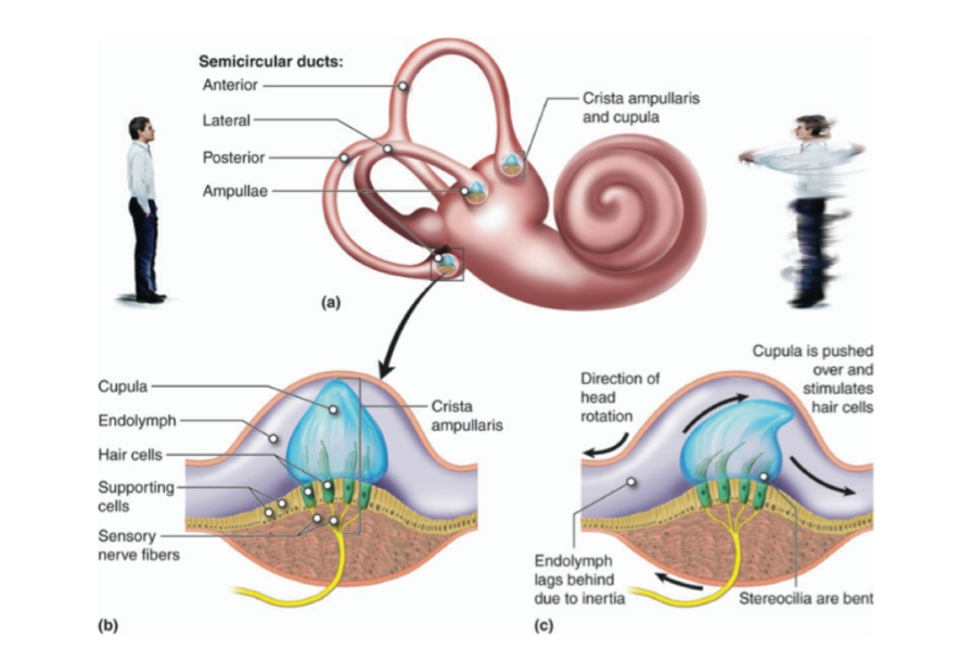
Q.39 Where do the semicircular ducts open?
Utricle.
Q.40 What is the blood supply of the internal ear?
Arterial supply:
By labyrinthine artery, which is a branch of anterior inferior cerebellar artery.
Also some branches from the stylomastoid artery which also supplies the middle ear.
Venous drainage:
Into superior petrosal or transverse sinus.
Q.41 What are the functions of the Internal ear?
Cochlear portion: Hearing
Vestibular part: Equilibrium.
Semicircular canals act as kinetic labyrinth while utricle and saccule as static labyrinth.
Q.42 What are the receptor cells for hearing and where they are located?
The receptors are neuroepithelial hair cells situated on the organ of Cort in the duct of the cochlea, just above the basilar membrane.
Q.43 What are the receptors for equilibrium and where they are located.
Receptor cells are hair cells located on the macula of utricle and saccule (for static balance) and on crista of ampulla of semicircular ducts (for kinetic balance).
Comments (0)