Orbit (Viva)
6 years ago 2225
Q.1 Name the different layers of the eyelid.
Q.2 What are the glands found in eyelid?
Q.3 What are the modifications of palpebral fascia?
Q.4 What is the nerve supply of eyelids?
Q.5 What is Chalazion?
It is chronic inflammation of the tarsal gland, causing localized swelling.
Q.6 What is Stye?
It is an infection of Zeis’s gland. The lid margin is oedematous and the gland is swollen and painful.
Q.7 What are the constituents of the lacrimal apparatus?
Q.8 What is the nature of the lacrimal gland?
Exocrine and serous.
Q.9 What are the parts of the lacrimal gland?
Q.9 Why the removal of palpebral part of the gland is equivalent to functional removal of the whole gland?
Because the ducts of the orbital part also pass through the palpebral part. So when palpebral part is removed the secretions of orbital part cannot be drained.
Q.10 What is the advantage of the blinking of lids?
It helps to spread the lacrimal fluid in front of the eye and deep surface of lids, thus keep the conjunctiva and cornea moist.
Q.11 What is the valve of Hasner?
It is a fold of mucous membrane at the lower end of the nasolacrimal duct.
Q.12 What is Epiphora?
Leakage of tears down the face, due to the blockade of the nasolacrimal duct.
Q.13 What is Tenon’s Sheath?
It is a thin membranous sheath around the eyeball. Extends from optic nerve to sclerocorneal junction. Eyeball can move freely within it.
Q.14 Name the structures piercing the fascial sheath of the eyeball.
Q.15 What is the suspensory ligament of Lockwood?
It is thickened Tenon’s capsule in the lower part. Formed by the union of margins of the sheath of inferior rectus and inferior oblique with medial and lateral check ligaments.
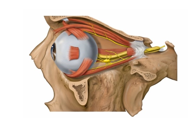
Q.16 Name the extra-ocular muscles?
Voluntary muscles:
Involuntary muscles:
Q.17 What is the origin of rectus muscles?
They arise from the corresponding part of the common tendinous ring which surrounds the optic canal and encloses a part of the superior orbital fissure.
Q.18 What is the origin of oblique muscles?
Superior oblique: Body of sphenoid above and medial to optic canal.
Inferior oblique: Anterior and medial part of the floor of orbit from maxilla just lateral to nasolacrimal groove.
Q.19 Which muscles produce the different movements of the eyeball?
| Upwards: Superior rectus, Inferior oblique |
| Downwards: Inferior rectus, Superior oblique |
| Inwards: Medial rectus, Superior rectus, Inferior rectus |
| Outwards: Lateral rectus, Superior oblique, Inferior oblique |
| Extorsion: Inferior oblique, Inferior rectus |
| Intorsion: Superior oblique, Superior rectus. |
Q.20 What is the nerve supply of extraocular muscles?
| Superior oblique: Trochlear nerve |
| Lateral rectus: Abducent nerve |
| Superior, inferior and medial rectus, inferior oblique and levator: Oculomotor nerve. |
| Involuntary muscles: By sympathetic fibers |
Q.21 What are conjugate movements of the eye?
The normal co-ordinated movements of both eyes are called conjugate movements. These are usually horizontal and vertical.
Q.22 What is Nystagmus?
It is involuntary rhythmical oscillatory movements of the eye due to incoordination of ocular muscles.
Q.23 What is Squint?
It is the abnormal deviation of the eye due to weakness or paralysis of a muscle.
Comments (0)