Pharynx (Viva)
6 years ago 4395
PHARYNX
Q.1 What is the length of pharynx?
12 cm
Q.2 What is the extent of pharynx?
Superiorly: Base of skull including posterior part of the body of sphenoid and basilar part of the occipital bone.
Inferiorly: C6 vertebra or lower border of the cricoid cartilage.
Q.3 What are the attachments of pharynx on each side?
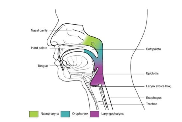
Q.4 What are the parts of pharynx?
Q.5 What are the characteristic features of nasopharynx?
Q.6 What are the features of the lateral wall of nasopharynx?
Q.7 What is the clinical importance of pharyngeal recess (Fossa of Rosenmuller)?
It forms a flat pocket. A catheter missing the tubal opening may enter recess and perforate the pharyngobasilar fascia and enter the ICA (Internal carotid artery).
Q.8 What structures form the junction of nasopharynx and oropharynx?
Q.9 What is Isthmus of fauces?
Also known as oropharyngeal isthmus and it represents the junction of the oropharynx and oral cavity.
Q.10 What are the boundaries of oropharyngeal isthmus?
Above: Soft palate
Below: Posterior part of tongue
On either side: Palatoglossal arches
Q.11 Which muscle helps in closure of oropharyngeal isthmus?
Palatoglossus.
Q.12 What is the extent of the laryngeal part of pharynx?
From third to sixth cervical vertebra.
Q.13 Where does the junction of oropharynx and laryngopharynx lie?
Upper border of the epiglottis
Q.14 What is the position of ‘piriform fossa’?
It is present on each side of the inlet of larynx. The fossa is bounded medially by aryepiglottic fold and laterally by thyroid cartilage and thyrohyoid membrane. Beneath mucosa of fossa lie internal laryngeal nerve.
Q.15 What is the clinical importance of piriform fossa?
A foreign body may lodge here. If removed, damage to the internal laryngeal nerve may occur leading to anesthesia in the supraglottic part of larynx. Thus, leading to aspiration pneumonia.
Q.16 What are the different layers forming the wall of pharynx?
Q.17 Name the muscles of the pharynx.
Longitudinal layer:
Circular layer:
Q.18 What is the nerve supply of the pharynx?
Motor fibers:
Sensory:
Glossopharyngeal and vagus.
Nasopharynx:
Maxillary nerve.
Taste:
Internal laryngeal branch of the vagus.
Secretomotor:
Greater petrosal nerve.
Q.19 What is pharyngeal plexus?
It is a plexus of nerve, present beneath the buccopharyngeal fascia.
Formed by:
Q.20 What is the arrangement of constrictors of pharynx?
These are arranged like three flower pots one above the other, the lower constrictor overlapping the upper one.
Q.21 What is the ‘sinus of Morgagni’?
It is a semilunar gap between the base of the skull and the upper border of superior constrictor.
Q.22 What are the structures passing through the sinus of Morgagni?
Q.23 What are the structures passing through the gap of superior and middle constrictor?
Q.24 Name the structures passing between middle and inferior constrictors.
Q.25 Name the structures passing through the gap between inferior constrictor and esophagus.
Q.26 What is Killian’s dehiscence?
This is a weak part in the posterior wall of pharynx, between thyropharyngeal and cricopharyngeal part of inferior constrictor.
Q.27 What is the clinical importance of Killian’s dehiscence?
Pharyngeal diverticula may be formed due to out-pouching at dehiscence.
Comments (0)