Tongue (Viva)
6 years ago 3529
TONGUE
Q.1 What are the parts of the tongue?
At the junction is a V-shaped groove with apex of V pointing backward sulcus terminalis, with a median pit, foramen caecum.
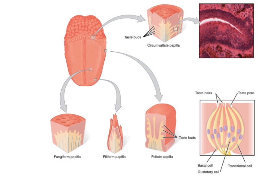
Q.2 What are papillae and what are their type?
Papillae are projections of mucous membranes situated in anterior 2/3 of the tongue on the dorsal surface.
Types:
Vallate papillae:
8-12 in number.
Situated immediately in front of the sulcus terminalis. Each is a cylindrical projection surrounded by a circular sulcus.
Fungiform papillae:
Numerous.
Near tip and margins of tongue. Each has a narrow pedicle and a large rounded head.
Filiform papillae:
Most numerous.
Covers presulcal area of the dorsum of the tongue, pointed and covered with keratin.
Foliate papillae:
On lateral margin of posterior past. Appear as vertical folds but are not true papillae.
Q.3 Name the muscles of the tongue.
Tongue is divided into two halves by a midline fibrous septum.
Each half has:
Q.4 What is the lymphatic drainage of the tongue?
Lymphatics from near midline can pass either to the left or right side lymph nodes.
Q.5 Why jugulo-omohyoid node is called ‘lymph node of tongue?
Because it drains most of the lymph from the tongue.
Q.6 What is the nerve supply of the tongue?
Motor nerves:
Sensory:
Special sensory.
Q.7 What are the functions of the tongue?
Q.8 Where the taste buds are situated?
No taste buds in filiform papillae.
Q.9 How the tongue is developed?
| Anterior 2/3: First branchial arch by two lingual swellings and one tuberculum impar. |
| Posterior 1/3: Third arch by cranial half of hypobranchial eminence. |
| Posterior part: Fourth arch. |
| Muscles: Occipital myotomes. |
| Connective tissue: Local mesenchyme |
Q.10 How the bleeding from the lacerated tongue is stopped?
By applying pressure posterior to the area of laceration, because the lingual artery supplying it runs forwards.
Q.11 What does foramen caecum represents?
The site of down growth of thyroglossal duct.
Q.12 What is the clinical importance of attachment of genioglossus to the genial tubercles of mandible?
In unconscious patients or during general anesthesia, the tongue may fall back and obstruct the respiratory passage. So, the advantage of this attachment is taken by pulling the mandible forwards which prevents the falling back of the tongue.
Q.13 What are the developmental anomalies of the tongue?
Comments (0)