Triangles of Neck (Viva)
6 years ago 4673
Q.1 Name the structures palpable anteriorly in the midline of the neck.
Q.2 What is Platysma?
It is a subcutaneous muscle forming a thin fleshy sheath running upwards and medially on the neck from deltoid and pectoral fasciae to the base of mandible. It is supplied by the cervical branch of the facial nerve.
Functions:
Q.3 What is ‘jugular arch’?
A transverse channel in the suprasternal space connecting the two anterior jugular veins.
Q.4 What is the position of ‘subhyoid bursa’? What is its function?
Position:
Between the posterior surface of body hyoid bone and thyrohyoid membrane.
Function:
Lessens friction between the above two structures during swallowing.
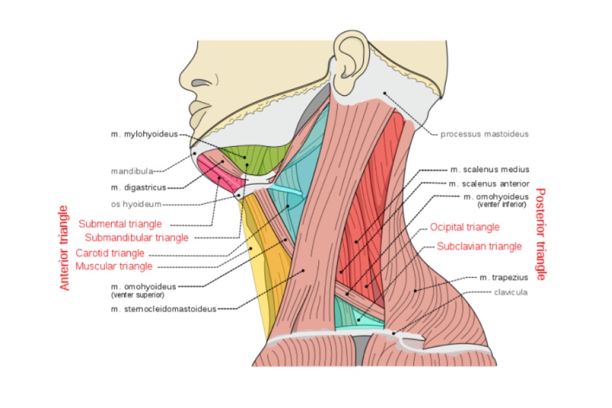
Q.5 What are the boundaries of the ‘anterior triangular’ of the neck?
Anterior: Anterior median line of neck.
Posterior: Anterior border of sternomastoid.
Base: Base of the mandible and a line joining angle of the mandible to the mastoid process.
Apex: Manubrium sterni.
Q.6 What are the subdivisions of the anterior triangle?
Q.7 What are the areas drained by submental lymph nodes?
Q.8 What are the areas drained by submandibular lymph nodes?
Q.9 What are the boundaries of the digastric triangle?
| Anteroinferiorly: Anterior belly of digastric |
| Posteroinferiorly: Posterior belly of digastric Stylohyoid |
| Base: Base of the mandible and a line joining angle of the mandible to the mastoid process |
| Roof: Skin Superficial fascia: Has platysma and cervical branch of facial nerve Deep fascia: Splits to enclose submandibular gland |
| Floor: Mylohyoid, Hyoglossus, and middle constrictor muscle. |
Q.10 Name the various structures lying over hyoglossus muscle.
Q.11 Name the various structures passing deep to hyoglossus muscle.
Q.12 Name the structures passing between external and internal carotid arteries.
Q.13 What are the boundaries of the carotid triangle?
| Superiorly: Posterior belly of digastric, Stylohyoid |
| Anteroinferiorly: Superior belly of omohyoid |
| Posteriorly: Anterior border of sternomastoid |
| Roof: Skin Superficial fascia having platysma, cervical branch of the facial nerve and transverse cutaneous nerve of neck Investing layer of deep fascia |
| Floor: Thyrohyoid, Hyoglossus, Middle constrictor, and Inferior constrictor. |
Q.14 Name the contents of the carotid triangle.
• Arteries:
Common carotid, Internal carotid, and External carotid and its branches.
• Veins:
Internal jugular, Common facial, Pharyngeal, Lingual and Superior thyroid.
• Nerves:
Vagus, Superior laryngeal, Hypoglossal and Sympathetic chain.
• Carotid sheath with its contents
• Deep cervical lymph nodes.
Q.15 What is the position, structure, and function of carotid sinus?
Position:
At the termination of the common carotid artery as slight dilatation.
Characteristics:
Media thin,
Adventitia thick,
Rich innervation by plexus formed by glossopharyngeal (mainly), sympathetic and vagus nerve.
Function:
As a baroreceptor helps to regulate blood pressure.
Q.16 What is the position, structure, and function of carotid bodies?
Position:
Present at the bifurcation of the common carotid artery near carotid sinus.
Structure:
Has characteristic glomus cells, similar to neurons.
Also has sympathetic and parasympathetic postganglionic neurons and afferent nerve terminals from the glossopharyngeal nerve.
Function:
Act as chemoreceptors to monitor oxygen and carbon dioxide levels in the blood by reflexly controlling the rate and depth of respiration.
Q.17 What are the branches of the external carotid artery?
Anteriorly:
Superior thyroid, Lingual and Facial.
Posteriorly:
Occipital and Posterior auricular.
Medial: Ascending pharyngeal.
Terminal:
Maxillary and Superficial temporal.
Q.18 Name the infrahyoid muscles.
Q.19 Name the suprahyoid muscles.
Q.20 What is the nerve supply of digastric muscle?
Anterior belly by mylohyoid branch of inferior alveolar nerve (V nerve) and posterior belly by the facial nerve.
Q.21 What are the attachments of omohyoid muscle?
It has two bellies:
Q.22 What are the boundaries submental triangle?
Base is formed by hyoid bone. Above and laterally on each side by the anterior belly of the digastric muscle. Floor by mylohyoid muscle. So half of it lies on each side of the midline of the neck.
Q.23 What are the boundaries of the muscular triangle?
Posterorinferiorly: Sternocleidomastoid muscle
Posterosuperiorly: Superior belly of omohyoid
Anteriorly: Anterior midline of neck.
Q.24 What are the contents of the muscular triangle?
Q.25 What are the boundaries of suboccipital trinagle?
Superomedially:
Rectus capitis posterior major, Rectus capitis posterior minor
Superolaterally:
Obliquus capitis superior
Inferiorly: Obliquus capitis inferior.
Roof:
Medially: Fibrous tissue
Laterally: Longissimus capitis
Floor:
Posterior arch of atlas
Posterior atlanto-occipital membrane.
Q.26 What are the contents of the sub-occipital triangle?
Q.27 What are the contents of ‘Suprasternal space of Burns’?
Q.28 What are the structures traversing supraclavicular space?
Q.29 What are the contents of the carotid sheath?
Q.30 Why the infections behind the prevertebral fascia do not extend to the posterior mediastinum?
Because the prevertebral fascia is attached to the fourth thoracic vertebra, which limits the downward spread of infection.
Q.31 What are the boundaries of the posterior triangle?
| Anterior: Posterior border of sternomastoid. |
| Posterior: Anterior border of trapezius. |
| Base: Middle 1/3 of clavicle. |
| Apex: Point where trapezius and sternomastoid meet. |
| Roof: Investing layer of deep cervical fascia. |
| Floor: Prevertebral layer of deep cervical fascia covering the muscles. |
Q.32 What are the structures present in the floor of the posterior triangle below deep cervical fascia?
Semispinalis capitis,
Splenius capitis
Levator scapulae,
Scalenus posterior,
Scalenus medius, and
Scalenus anterior.
By inferior belly of omohyoid in lower part, it is divided into the upper part (occipital triangle) and lower part (supraclavicular triangle)
Q.33 What are the contents of the posterior triangle of the neck?
Q.34 What are ‘Signal nodes’?
These are lymph nodes that are enlarged in the malignant growths of distant places e.g., left supraclavicular nodes in malignancy of stomach, testes, and other abdominal organs.
Q.35 What is the origin of sternomastoid?
Q.36 What is the nerve supply of sternomastoid?
Motor:
Spinal accessory nerve.
Sensory:
Ventral rami of C2,3.
Q.37 What is the action of sternocleidomastoid muscle?
When muscle of one side contacts, the head is tilted to the same side, and the face is rotated to the opposite side. When muscles of both sides act together, the head and neck are flexed.
Q.38 What is ‘torticollis’?
Also known as Wryneck. The head is bent to one side and chin points to the other side.
Q.39 How torticollis occurs?
It occurs due to the spasm of muscles supplied by spinal accessory nerve i.e., sternomastoid and trapezius.
Comments (0)