Major Blood Vessels of Thorax Region (VIva)
6 years ago 4254
Q.1 How superior vena cava is formed?
By the union of two brachiocephalic veins behind the lower border of first costal cartilage close to the sternum.
Q.2 Name the tributaries of superior vena cava.
Q.3 What is the pathway for the collateral circulation in obstruction of superior vena cava?
Q.4 How superior vena cava is developed?
PULMONARY TRUNK AND ARTERIES
Q.1 What is the course of pulmonary trunk?
It begins opposite the sternal end of the left 3rd costal cartilage and the upper end lies in front of the fifth thoracic vertebra. The bifurcation of the pulmonary trunk lies below the arch of the aorta.
Q.2 What are the relations of the right pulmonary artery?
Anterior:
Ascending aorta, Superior vena cava, and Upper right pulmonary vein.
Posterior:
Oesophagus and Right bronchus.
Q.3 What are the relations of the left pulmonary artery?
Posterior:
Left bronchus and Descending aorta.
Superiorly:
Connected to the arch of the aorta by ligamentum arteriosum.
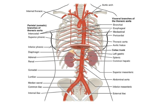
AORTA
Q.1 What are the parts of aorta in thorax?
Q.2 What is the course of ascending aorta?
It begins at the level of the lower border of 3rd costal cartilage behind the left half of the sternum. It runs upwards, forwards and to right, and continues as the arch of the aorta at sternal end of upper border of second right costal cartilage.
Q.3 What is Aortic sinus?
It is dilatation of the vessel wall at the root of the aorta above each cusp of the aortic valve.
Q.4 Name the branches of ascending aorta?
Q.5 What is the level of beginning and termination of the arch of aorta?
It begins behind the upper border of 2nd right sternochondral joint (lower border of T4) and ends at a lower border of the body of the 4th thoracic vertebra on the left side. Thus it begins and ends at the same level but it begins anteriorly and ends posteriorly.
Q.6 What are the posterior and to the right relations of the arch of aorta?
From behind forwards these are:
Q.7 Name the branches of arch of aorta.
Q.8 Name branches of descending thoracic aorta.
Q.9 What is aortic aneurysm?
It is a localized abnormal dilatation of aorta.
Q.10 What is the coarctation of the aorta?
It is the narrowing of the aorta, occurring usually immediately beyond the origin of the left subclavian artery. It leads to hypertension above the narrowing e.g., arms, neck, and head and hypotension below e.g., lower limb.
Q.11 What is the developmental origin of the aorta?
Q.12 What is ‘ductus arteriosus’?
It is communication present in fetal life connecting the left pulmonary artery with aorta just distal to the origin of the left subclavian artery. After birth, it gets obliterated and forms ligamentum arteriosum.
Q.13 What will happen if ductus arteriosus remains patent?
It causes progressive enlargement of the left ventricle and pulmonary hypertension.
AZYGOUS AND HEMIZYGOUS VEINS
Q.1 How azygous vein is formed and terminates?
By the union of right ascending lumbar and right subcostal vein at the level of T12 vertebra and terminates at the level of T4 vertebra into superior vena cava.
Q.2 Name the tributaries of azygous vein.
Q.3 Name the tributaries of hemiazygous vein.
THORACIC DUCT
Q.1 What is the length of the thoracic duct?
40 cm
Q.2 What is the extent of the thoracic duct?
Begins from Cisterna chyli near the lower border of the T12 vertebra. Ends into the angle of the junction between left subclavian and left internal jugular vein at the level of T2 vertebra.
Q.3 What are the relations of the thoracic duct in the aortic opening of the diaphragm?
Q.4 Name the tributaries of the thoracic duct.
In thorax:
At root of neck:
Q.5 From which areas the thoracic duct drains lymph?
Comments (0)