Trachea & Bronchi Anatomy
6 years ago 3789
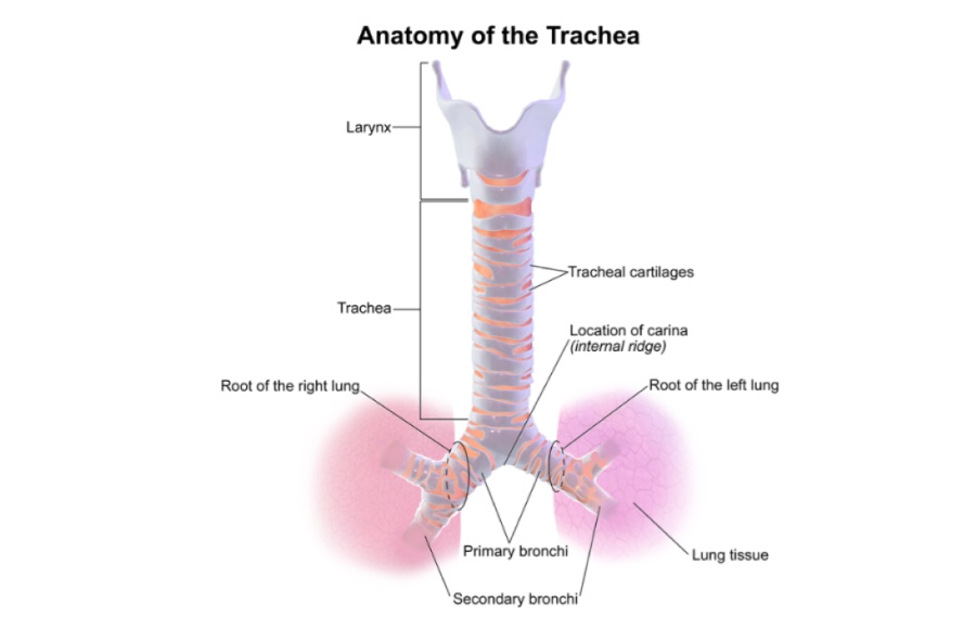
TRACHEA
Q.1 What is the extent of the trachea?
Trachea extends from 6th cervical vertebra (lower border of cricoid cartilage) to lower border of 4th thoracic vertebra where it divides into right and left bronchi.
Q.2 What are the relations of the cervical part of the trachea?
Anteriorly:
Posteriorly:
On each side:
Q.3 What are the relations of the thoracic part of the trachea.
Anterior:
Posterior:
Right side:
Left side:
Q.4 What is the arterial supply of trachea?
Inferior thyroid arteries
Q.5 What is the nerve supply of the trachea?
Parasympathetic nerves:
Vagus through recurrent laryngeal.
It is
• Sensory
• Secretomotor
• Motor to tracheal muscle
Sympathetic nerves:
Through middle cervical ganglion.
It is vasomotor.
Q.6 What is the lymphatic drainage of the trachea?
To
Q.7 What are the variations in the level of bifurcation of trachea with respiration?
Bifurcation of trachea, normally: Between T4 and T5 vertebra.
In deep inspiration: T6 vertebra.
In expiration: T4 vertebra.
Q.8 Where trachea can be palpated?
In suprasternal notch midway between sternal ends of two clavicles.
Q.9 What is ‘Tracheal tug’?
Arch of aorta lies in close relation to the trachea and left bronchus.
In the aneurysm of the aortic arch, a pull or drag is felt on the trachea which is known as ‘tracheal tug’.
Q.10 How the trachea appear in an X-ray?
Since the trachea is more radiolucent (because of air in it) than neighboring structures, it appears as a dark area passing downwards, backward and slightly to the right.
Q.11 In what conditions the tracheostomy is done?
Q.12 What is the commonest site for tracheostomy?
It is most commonly done in retrothyroid region after cutting the isthmus of the thyroid gland. Usually, the second and third tracheal rings are cut.
Q.13 Why the tracheostomy is difficult and dangerous in children?
Because
Q.14 What is the histological structure of the trachea?
Trachea consists of following layers from within outwards:
Q.15 What is the advantage of the posterior gap in tracheal cartilage?
Posteriorly, the esophagus lies close to the trachea so the esophagus can dilate into the posterior membranous part, during the passage of food bolus.
BRONCHI
Q.1 What are the differences between right and left main bronchus?
| Right main bronchus | Left main bronchus |
| Wider, shorter (2.5 cm) more vertical | Narrower, longer (5 cm), and less vertical |
| Passes to root of lung at T5 | Passes to root of lung at T6 |
| Divides into 3 lobar bronchi | Divides into 2 lobar bronchi |
| Relations: | Relations: |
| – Azygos vein arches over it from behind to reach superior vena cava | It passes below the arch of aorta, in front of esophagus and descending aorta |
| – Right pulmonary artery lies first below and then anterior to it. | Left pulmonary artery lies first anterior and then above it. |
| – Right upper pulmonary vein covers right principal bronchus | Left upper pulmonary vein crosses in front of the bronchus. |
Q.2 Why the foreign bodies and aspirated material tend to pass into the right bronchus rather than into left?
Because of the greater width and more vertical course of the right bronchus.
Also read: Anatomy Question Collection
Also read: Anatomy Questions & Answers
Also read: Anatomy notes
Comments (0)