Thoracic Cage & Vertebrae, Sternum (Viva)
6 years ago 4560
THORACIC CAGE
Q.1 How thoracic cage is formed?
Anteriorly: Sternum
Posteriorly: Twelve thoracic vertebrae and intervertebral discs
One each side: Twelve ribs with their cartilages
Q.2 What variations occur in thorax with age?
Q.3 What are the boundaries of ‘thoracic inlet’?
Q.4 What is the direction of the plane of the inlet of the thorax?
Downwards and forwards with an obliquity of about 45 degrees. The upper border of manubrium sterni lies at the level of the upper border of the T3 vertebra.
Q.5 What is Simson’s fascia and what are its attachments?
It is a triangular membrane at the thoracic inlet (Diaphragm of inlet of the thorax).
Attachments:
Apex: Tip of transverse process of C7 vertebra.
Base: laser border of the first rib and its cartilage.
Inferior surface: Fused with cervical pleura.
Q.6 Name the structures passing through thoracic inlet.
Muscles:
Arteries:
Nerves:
Veins:
Others:
Q.7 What are the boundaries of the outlet of the thorax?
THORACIC VERTEBRAE
Q.1 What are the typical’ and ‘atypical’ thoracic vertebrae?
Typical thoracic vertebrae: 2nd to 8th, they have common features.
Atypical vertebrae: 1st, 9th to 12th, they have special features.
Q.2 What are the structures attached to the transverse process?
| Lateral costotransverse ligament: At tip. |
| Inferior costotransverse ligament: On anterior surface. |
| Superior costotransverse ligament: On lower border. |
| Intertransverse muscles: Upper and lower borders. |
| Levatorcostae: To posterior surface. |
Q.3 What is the characteristic feature of a typical thoracic vertebrae?
It has costal facets on sides of vertebral bodies and transverse processes for articulation with ribs.
Q.4 What movement is possible in the thoracic spine?
Rotation, greater in the lower thoracic region as compared to the upper thoracic region.
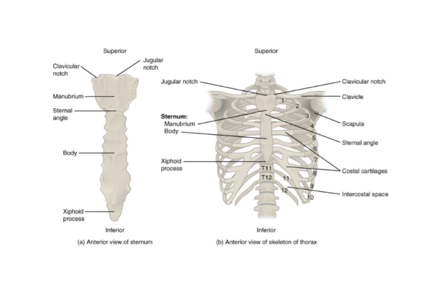
STERNUM
Q.1 What are the parts of the sternum?
Q.2 What is Jugular notch?
Also called suprasternal notch, present in the middle of the superior border of the manubrium.
Q.3 What is the level of jugular notch?
It lies at the level of the intervertebral disc between T2 and T3 vertebra.
Q.4 What is the sternal angle (Angle of Louis)?
It is the angle formed at the junction of manubrium and body of the sternum. It is convex forwards.
Q.5 What is the level of sternal angle?
Intervertebral disc between T4 and T5 vertebra.
Q.6 What is the clinical significance of the sternal angle?
It is an important landmark for counting ribs as 2nd costal cartilages articulate with sternum of this level.
Q.7 Name the structures lying at the level of the sternal angle.
Q.8 Why the sternum is commonly used for getting a specimen of bone marrow?
Because the cortical bone of sternum is very thin and subcutaneous. It is therefore easily accessible.
Q.9 What types of joints are present between different parts of the sternum?
Between manubrium and body (Manubriosternal joint):
Secondary cartilaginous joint.
Between body and xiphisternum (Xiphisternal joint):
Primary cartilaginous joint
Q.10 Which rib is attached to junction of the body with the xiphoid process
Seventh costal cartilage
Q.11 What type of joint is the sternocostal joint?
Synovial joint
Q.12 What is the level of Xiphisternal joint?
9th thoracic vertebra.
Q.13 What is ‘Funnel chest’?
Deformity of the chest in which the body of the sternum and xiphoid process is depressed. This predisposes to respiratory and cardiovascular disturbances.
Q.14 What is ‘Pigeon chest’?
It is a condition in which deformity of chest occurs due to forward projection of sternum and flattening of the chest on either side.
Q.15 What is ‘Ectopia cordis’?
In this, the sternum and adjoining parts of costal cartilages and ribs are missing. So that the heart can be seen from outside.
Also read: Anatomy Question Collection
Also read: Anatomy Questions & Answers
Also read: Anatomy notes
Comments (0)