Diaphragm (Viva)
6 years ago 6729
Q.1 What is the origin of diaphragm?
Arise from the periphery, in three parts:
Q.2 What are lumbocostal arches?
These are tendinous arches in the fascia covering the muscles in the posterior abdominal wall, e.g. medial lumbocostal arch (medial arcuate ligament) in fascia over the upper part of psoas major and lateral lumbocostal arch (lateral arcuate ligament) in fascia over the upper part of quadratus lumborum.
Q.3 What is the origin of the crus of the diaphragm?
Right crus: From anterolateral surface of body of L1,2,3.
Left crus: From anterolateral surface of body of L1,2.
The medial margins of two crura join to form the median arcuate ligament.
Q.4 What is the insertion of muscle fibers of the diaphragm?
Trilobed central tendon, which lies below and is fused to the pericardium.
Q.5 What is the nerve supply of diaphragm?
Motor: Phrenic nerve (C3,4).
Sensory:
– Phrenic nerves: Central part.
– Lower six thoracic nerves: Peripheral part.
Q.6 What are the other structures supplied by the phrenic nerve?
Sensory fibers to:
Q.7 What are the functions of the diaphragm?
Q.8 What are the variations in the position of the diaphragm with posture?
Level of diaphragm is:
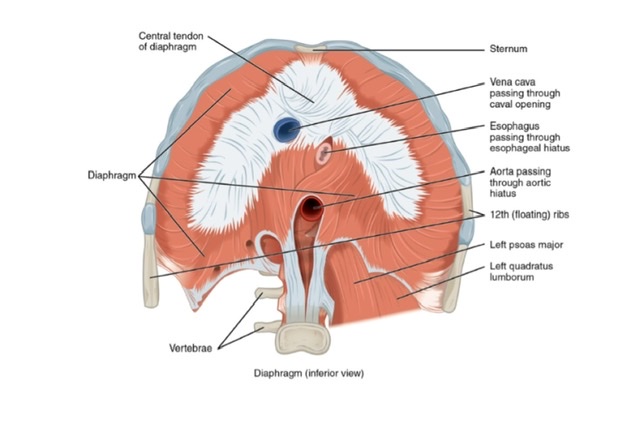
Q.9 Name the structures passing through the opening of the diaphragm.
Q.10 Why irritation of diaphragm causes pain in shoulder tip?
Because phrenic nerve and supraclavicular nerves have the same root value, i.e. C3,4.
Q.11 What is ‘eventration’ of diaphragm?
This is a congenital defect, in which the high position of the diaphragm occurs due to the replacement of the left half of the diaphragm by a fibrous membrane.
Q.12 What is foramen of Morgagni?
Also called space of Larry. It is a space between the xiphoid and the costal origins of diaphragm.
Site of congenital hernia. More common on the right side.
Q.13 What is foramen of Bochdalek?
This is the commonest site of congenital diaphragmatic defects in the periphery of the diaphragm in the region of the 10th and 11th ribs attachment.
Defect is in the posterolateral part of the dome on the left side of the diaphragm resulting from the failure of closure of pericardiopleural canal.
Q.14 What is the development origin of diaphragm?
Diaphragm is developed from:
Q.15 What is the diaphragm? What are its attachments?
The diaphragm is a large muscle which forms a partition between the cavities of the thorax and abdomen origin.
The origin of the diaphragm can be divided into sternal, costal, and lumbar vertebral parts.
The sternal part consists of two slips: right and left which arise from the back of the xiphoid process. The costal part consists of broad slips one from the inner surface of each of the lower six ribs (7th to 12th) and their costal cartilages.
The lumbar part consists of two crura (right and left) that arise from the anterolateral aspects of the bodies of lumbar vertebrae and of fibers that arise (on either side) from tendinous arches called the lateral and medial arcuate ligaments. The right crus is larger than the left; it crusis from the bodies of vertebrae L1, L2, and L3 and from the intervening intervertebral discs. The left crus similarly arises from vertebrae L1 and L2.
Insertion:
From its extensive origin, the muscular fibers of the diaphragm run upwards and converge to the inserted on the margins of a large, flat, central tendon, which is located just below the pericardium and heart.
Q.255 Describe the apertures present in the diaphragm.
The apertures present in the diaphragm are as follows
Also read: Anatomy Question Collection
Also read: Anatomy Questions & Answers
Also read: Anatomy notes
Comments (0)