NERVES, MUSCLES, FASCIA AND ARTERY OF PELVIS (Viva)
6 years ago 3001
Q.1 Name the branches of the internal iliac artery.
In males:
In females:
Same as above except the inferior vesical is replaced by the vaginal artery. Also uterine artery.
Q.2 Name the tributaries of the internal iliac vein.
Veins arising outside the pelvic wall:
Veins arising from venous plexuses of pelvic viscera:
Q.3 How sacral plexus is formed?
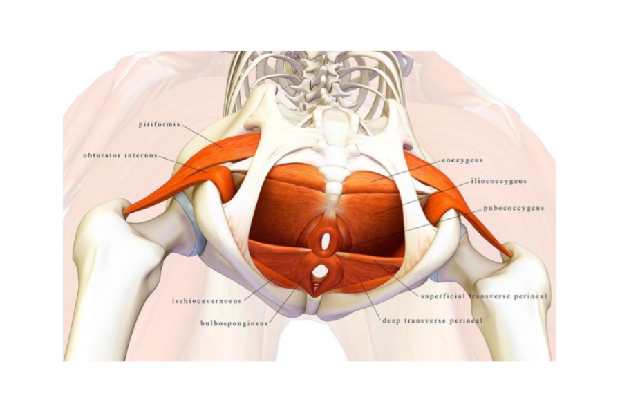
Q.4 Describe the levator ani muscle.
The levator ani is a brood-thin muscle that is situated on the side of the pelvis and supports the viscera in the pelvic cavity.
Origin:
The L. ani muscle originates from the following structures (front to back)
– Pelvic surface of the body of the pubis
– Obturator fascia
– Spine of ischium
Insertion
– The anterior-most fibers pass across the sides of the prostate (in males) and sides of the vagina (in females) to end into the perineal body.
– The intermediate fibers pass across the sides of the rectum and become continuous with these of the opposite side side behind the anorectal junction.
– The posterior-most fibers of lateral ani are attached to the coccyx and to a fibrous band called the anococcygeal ligament. The posterior margin of the muscle is continuous with the coccyx.
Q.5 What are the parts of levator ani?
– Anterior fibers from levator prostate in males and pubovaginalis in females.
– Middle fibers form puborectalis.
– Posterior fibers form pubococcygeus properly.
Q.6 What is the insertion of levator ani?
Perineal body
Q.7 Where the pain of pelvic organs is referred to?
The autonomic supply which is sensory to pelvic organs is by S2,3,4 spinal segments, which also gives cutaneous nerves to the perineum. Therefore disease of pelvic organs causes referred pain in the perineum.
Q.8 What is the hiatus of Schwalbe and what is its clinical importance?
Levator ani arises from the pubic bone in front, ischial spine behind, and obturator fascia between these points. Sometimes, it arises from a tendinous sling, which is attached to the bone in front and behind and not to the fascia at all. Thus, a potential gap exists between the sling and obturator fascia, called the hiatus of Schwalbe.
Clinical importance:
The pelvic peritoneum may herniate through it into the ischiorectal fossa.
Comments (0)