Osteology of Abdomen & Pelvis (Viva)
6 years ago 4411
Q.1 How the sacrum is formed?
By the fusion of 5 sacral vertebrae.
Q.2 What is the anatomical position of the sacrum in the body?
Q.3 What are the relations and attachments of the ala of sacrum?
Q.4 What are the relations of the pelvic surface of the sacrum?
Q.5 How ‘Sacral hiatus’ is formed?
By failure of fusion of laminae of S5 vertebrae posteriorly.
Q.6 Name the structures emerging at the sacral hiatus.
Q.7 What is the origin of erector spinae?
It has a linear U-shaped origin from the dorsal aspect of the sacrum. The medial limb of the U is attached to the spinous tubercles and the lateral limb to the transverse tubercles.
Q.8 How will you identify the lumbar vertebra?
Q.9 Name the structures attached to the spine of the lumbar vertebra.
Q.10 What is ‘Sacralisation of lumbar vertebra’?
Fusion of the L5 vertebra or its transverse process on one or both sides with the sacrum.
Q.11 What is ‘spina Bifida?
Failure of the posterior fusion of two halves of the neural arch with each other results in a bony gap. Through the gap, the meninges and spinal cord may herniate out in the mid-line.
Q.12 How will you differentiate between male and female sacrum?
| Male sacrum | Female sacrum | |
| Length and breadth | Longer and narrower | Shorter and wider |
| Body and ala | Transverse diameter of the body of S1 is larger than that of ala | The transverse diameter of the body is of S1 equal to the width of the ala |
| Auricular surface | Dorsal concavity is less marked | Dorsal concavity is more marked. |
| Pelvic curve | Sacrum more uniformly curved, and concavity is shallower | Curvature is irregular, concavity is deeper. |
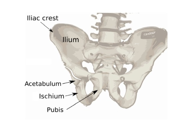
BONY PELVIS
Q.1 How the bony pelvis is placed in anatomical position?
Q.2 What are the parts of the pelvis?
The pelvis is divided by the pelvic brim (pelvic inlet) into two parts:
Q.3 What are the boundaries of the pelvic inlet (superior pelvic aperture)?
Anteriorly: Upper margin of the pubic symphysis.
Posteriorly: Sacral promontory.
On each side:
Anterior margin of the ala of sacrum, and linea terminales which includes an arcuate line of the ilium, pectineal line of the pubis, and pubic crest.
Q.4 What are the boundaries of the inferior pelvic aperture?
Anteriorly: Pubic arch
Posteriorly: Lateral margin of sacrum and coccyx
On each side:
Ischial tuberosity, lesser sciatic notch, ischial spine, and greater sciatic notch.
The lateral margin is formed by the sacrotuberous ligament.
Q.5 What is the axis of the pelvis?
This is an imaginary line joining the central points of anteroposterior diameters of the pelvic outlet and inlet. It is J-shaped, directed first downwards and backward and then downwards and forwards.
Q.6 How will you differentiate between male and female pelvis?
| Male pelvis | Female pelvis | |
| General Features | Heavier and stronger | Lighter and thinner |
| Bony markings: More prominent |
Less prominent. | |
| Ilia | Less vertical | More vertical |
| Greater in height | Lesser in height | |
| Iliac fossae: | Deeper | Shallower |
| Intercristal diameter: | Greater | Smaller |
| Pelvic inlet | Smaller and heart-shaped. | Larger and more circular. |
| Body of S1 vertebra forms: | Pubic tubercles are nearer because the pubic crest is narrower. | Pubic tubercles are wider apart because the pubic crest is longer. |
| More than the width of the lateral part | Equal to the width of the lateral part | |
| Pelvic outlet | Smaller | Larger |
| Sub-pubic angle: | 50-60° | 80-85° |
| Ischial tuberosities: | Less everted | More everted |
| Coccyx: | Less vertical | More vertical |
| Sciatic notches | ||
| Greater sciatic notch: | Narrower | Wider |
| Ischial spines: | Closer and inturned | Wider apart |
| Pelvic walls | ||
| Concavity of sacrum: | Shallower | Deeper |
| Sacrum: | Long and narrow | Short and wide |
| Obturator foramen: | Larger and ovoid | Smaller and triangular |
| Acetabulum: | Larger and faces fewer forwards | Smaller and faces more forwards |
| Puboischial index: | < 90 | > 90 |
| Pelvic cavity | Longer and more conical | Shorter and cylindrical |
Q.7 What is the diagonal conjugate of the pelvis?
The diagonal conjugate is an anteroposterior diameter between the sacral promontory and pubic symphysis. This is normally at least 11.5 cm.
Q.8 What is the normal shape of the female pelvis and what are the different variations in it?
JOINTS OF PELVIS
Q.1 Name the joints of the pelvis
Q.2 Name the ligaments of the sacrococcygeal joint.
Q.3 What variety of joint pubic symphysis is?
Secondary cartilaginous joint
Q.4 Name the ligaments of the sacroiliac joint.
Comments (0)