Portal Vein, Coeliac & Superior Mesenteric vessels (VIVA)
6 years ago 6656
PORTAL VEIN
Q.1 What is the characteristic feature of the portal vein?
Portal vein is one vein that begins and also ends in capillaries, i.e. the vein formed from capillaries of an organ, enter another organ where they divide into another set of capillaries.
Q.2 Name the areas from which the blood is drained by the portal vein.
Q.3 What are the divisions of the portal vein?
The portal vein is divided into 3 parts: Intraduodenal, gastroduodenal, and supraduodenal.
Q.4 What are the relations of different parts of the portal vein?
Q.5 How does the portal vein form and terminate?
Formation:
The portal vein is formed at the level of the L2 vertebra behind the neck of the pancreas, by the union of superior mesenteric and splenic veins.
Termination:
It ends at the right end of porta hepatis by dividing it into a right and a left branch.
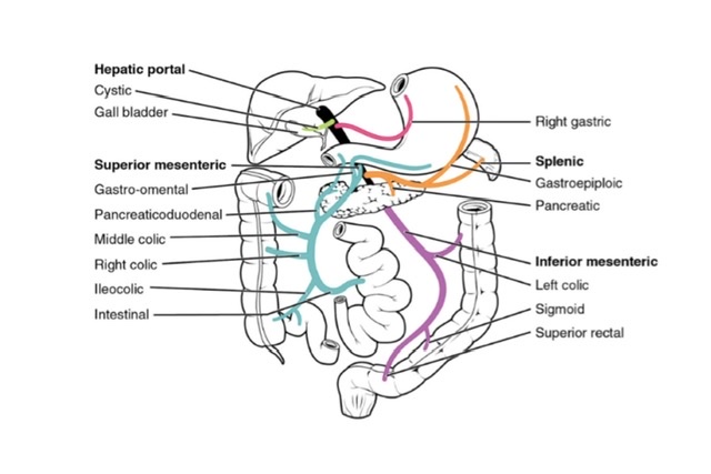
Q.6 What are the tributaries of the portal vein?
Q.7 What are the sites of portal-systemic communications? The name also the portal and systemic veins forming these.
| Sites | Portal vein | Systemic vein |
| Umbilicus | The left branch of the portal vein through a paraumbilical vein | Veins of the anterior abdominal wall |
| The lower end of the esophagus | Oesophageal tributaries of left gastric vein | Oesophageal tributaries of the accessory hemiazygous vein |
| Anal canal | Superior rectal vein | Middle and inferior rectal vein |
| Bare area of liver | Hepatic vein | Phrenic and intercostal veins |
| Posterior abdominal wall | Veins of the duodenum, ascending and descending colon | Retroperitoneal veins of the posterior abdominal wall (renal, lumbar, and phrenic veins) |
| Liver | Rarely ductus venosus remains patent and then connects the left branch of the portal vein | Inferior vena cava |
Q.8 What is the importance of portal-systemic communications?
These communications form the important pathways of collateral circulation in portal obstruction and portal hypertension.
Q.9 What is the developmental origin of the portal vein?
COELIAC TRUNK
Q.1 Name the structures supplied by the coeliac trunk.
The coeliac trunk supplies derivatives of foregut, i.e.
Q.2 How the coeliac trunk develops?
The coeliac trunk develops from one of the vitelline arteries (C7 segment).
Q.3 What are the branches of the coeliac trunk?
Q.4 What are the branches of the hepatic artery?
Q.5 What are the branches of the splenic artery?
SUPERIOR MESENTERIC VESSELS
Q.1 Name the structures supplied by the superior mesenteric artery.
The superior mesenteric artery supplies the derivatives of the midgut, i.e.
Q.2 At what level does the superior mesenteric artery arise?
It arises from the abdominal aorta at the L1 vertebra behind the body of the pancreas.
Q.3 Name the branches of the superior mesenteric artery.
Q.4 What are the relations of the superior mesenteric artery?
– Anteriorly:
Body of pancreas and splenic vein
– Posteriorly:
Aorta left renal vein,
Uncinate process of the pancreas, and
The third part of the duodenum
– To the right:
Superior mesenteric vein.
– It crosses:
Inferior vena cava
Right ureter
Right, psoas.
– To its right: |
Superior mesenteric vein
Q.5 What are the tributaries of superior mesenteric vein?
INFERIOR MESENTERIC VESSELS
Q.1 What are the structures supplied by the inferior mesenteric artery?
It supplies the derivatives of the hindgut, i.e.
Q.2 What are the branches of the inferior mesenteric artery?
Q.3 What is the ‘Marginal artery?
It is an arterial arcade situated along the concavity of the colon formed by anastomosis between ileocolic, right colic, middle colic, left colic and sigmoid arteries. Vasa recta arise from the marginal artery and supply the colon.
Q.4 What is the clinical importance of the marginal artery?
It forms extensive anastomosis, so it is capable of supplying the colon even in the absence of one of the main feeding trunks. This fact is utilized in the surgery of the colon.
Comments (0)